
Custom Total Ankle Replacement Using a Patient-Specific 3D-Printed Implant
A 48-year-old female with advanced osteoarthritis of the left ankle, where complex biomechanics and unique anatomy made conventional implant reconstruction difficult.
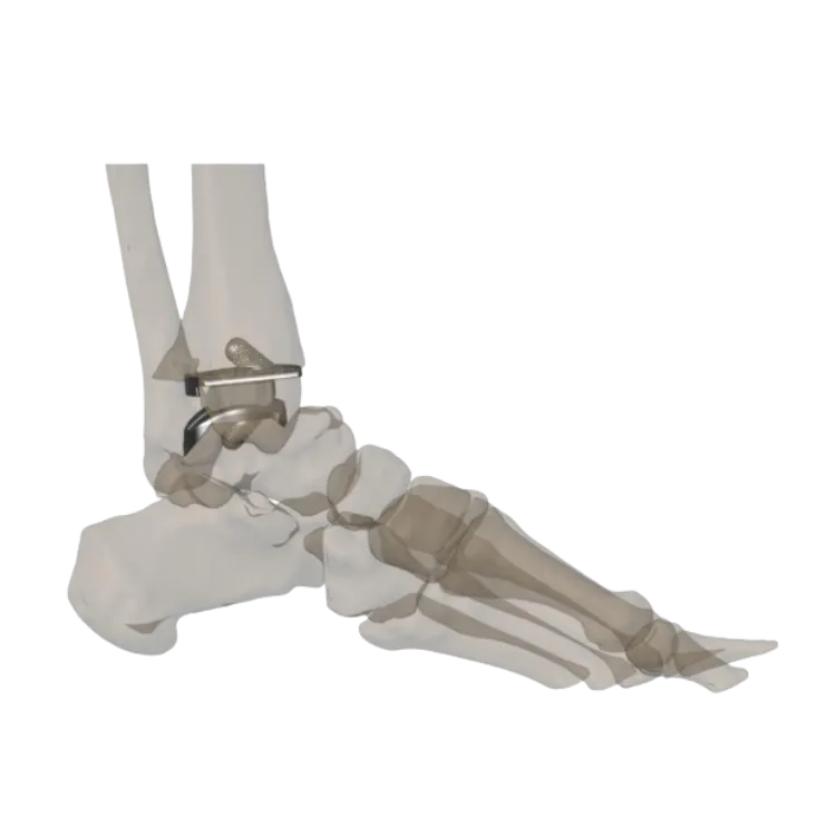
Patient overview
A 48-year-old female patient presented with advanced osteoarthritis of the left ankle joint, experiencing persistent pain, stiffness, and difficulty in daily movement due to severe cartilage degeneration.
Progressive joint misalignment had significantly impaired function and quality of life.
Clinical problem
The patient’s ankle biomechanics and individual anatomy made reconstruction with a conventional implant difficult. The degree of joint degradation meant a standard system could not be expected to restore alignment or distribute load predictably.
Preoperative planning
A successful ankle replacement depends heavily on restoring alignment, stability, and natural joint mechanics.
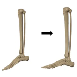
CT-based 3D segmentation was used to build an accurate anatomical model of the ankle joint. The foot was digitally repositioned into dorsiflexion to restore natural standing alignment, critical anatomical landmarks were established, and a personalised reconstruction strategy was agreed before any component was fabricated.
Patient-specific implant design
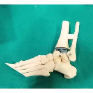
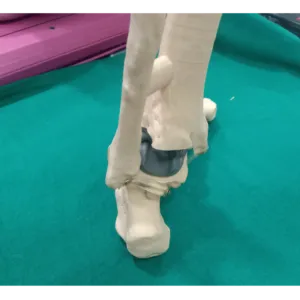
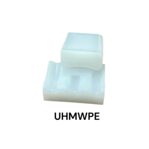
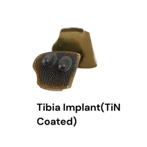
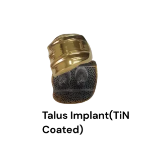
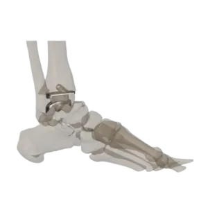
The customised ankle replacement system consisted of three components: a tibial component, a talar component, and a polyethylene insert.
The tibial and talar components were manufactured using advanced metal 3D printing technology with medical-grade titanium alloy (Ti6Al4V). The insert is UHMWPE, available in multiple thicknesses. Design features included a mobile-bearing configuration, peg fixation, a Ticellium surface for osseointegration and a TiN-coated articulating surface.
Surgical execution
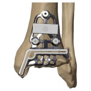
The plan was carried into theatre through patient-specific instrumentation, reproducing the planned tibial and talar resections and component positions directly on the patient’s anatomy.
Surgical outcome
The customised total ankle replacement was successfully performed by Dr. Harshvardhan Hegde.
Postoperative assessment demonstrated accurate restoration of ankle alignment, stable implant fixation, balanced load distribution and improved functional recovery.
Clinical significance
Where anatomy and biomechanics fall outside what standard total ankle systems accommodate, a fully patient-specific construct planned from CT data can restore alignment and function that would otherwise be difficult to achieve.
Related cases
 Foot & ankle
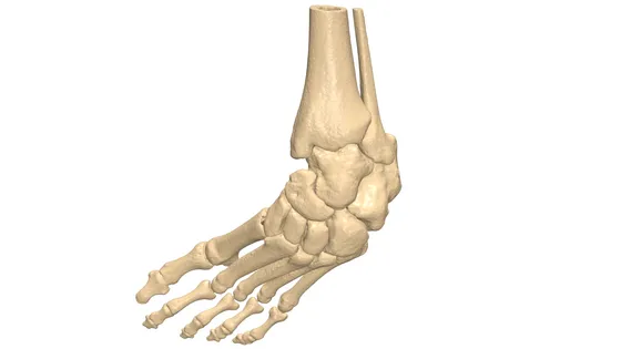
Club Foot Virtual Planning
Virtual osteotomy sequence and pre-op model for congenital talipes equinovarus
Read the case →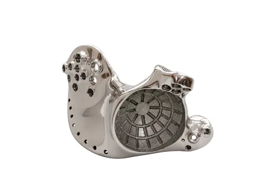 Oncology
Patient-Specific 3D Printed Hemipelvic Reconstruction with Constrained THA
Type I + II internal hemipelvectomy for Ewing's sarcoma
Read the case →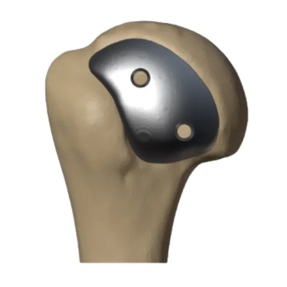 Shoulder
Custom Hill-Sachs Implant for Reconstruction of a Large Engaging Humeral Head Lesion
Patient-specific implant and drilling guide for a 22 × 41 mm humeral head defect
Read the case →Have a case like this?
Send the imaging and the clinical question — first 3D visualisation back within 24 hours.