
Patient-Specific Medial Malleolus Reconstruction Using a Custom 3D-Printed Titanium Implant
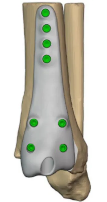
Case study: patient-specific 3D-printed titanium medial malleolus implant for ankle fracture-dislocation with bone loss, designed by Jajal Medical.
Case Overview
Medial malleolus fractures are significant ankle injuries that can compromise joint stability and articular congruity, particularly when associated with bone loss. Successful medial malleolus reconstruction requires anatomical restoration and stable fixation to support functional recovery.
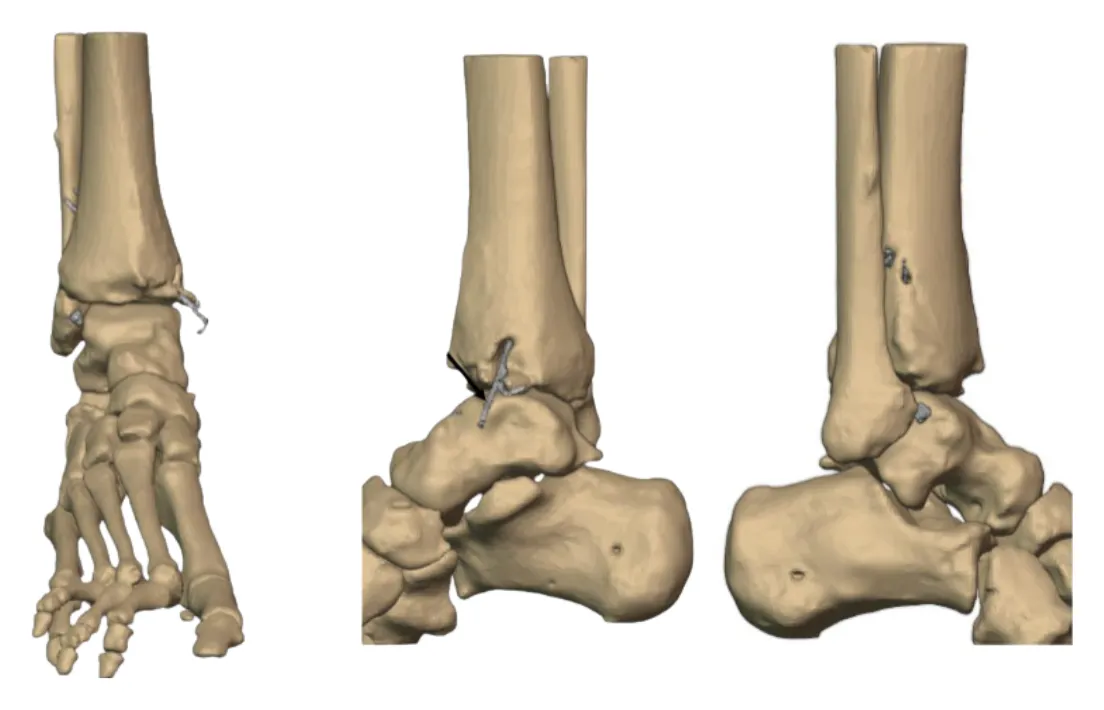
This case presents a 19-year-old male with a traumatic right ankle injury resulting in an open ankle fracture-dislocation and loss of the medial malleolus. A patient-specific medial malleolus implant was designed and manufactured in titanium to reconstruct the missing anatomical structure and restore ankle stability.
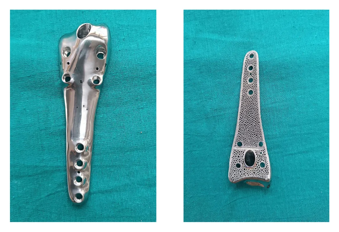
The custom titanium implant incorporated a polished surface for soft-tissue interaction and a 3D Printed Ticellium porous structure from Jajal Medical at the bone-contact interface to support osseointegration.
Clinical Background
The medial malleolus plays an essential role in maintaining ankle joint stability through its osseous structure and attachment to the deltoid ligament. Its loss or structural compromise can lead to ankle instability, altered joint mechanics, and long-term complications.
While conventional fixation methods, including lag screws, tension band wiring, and plates, are commonly used for medial malleolus fractures, extensive bone loss may require a customized reconstructive approach.
Patient-specific implants offer an alternative solution by allowing reconstruction based on the patient’s individual anatomy.
Patient-Specific Implant Design and Development
A CT scan of the patient’s lower extremity was acquired to obtain detailed anatomical data. The digital workflow for this custom ankle implant involved the following steps:
Step 1: Medical Image Segmentation
CT data were processed using MySegmenter (Jajal Medical Services Pvt. Ltd., India) to reconstruct the patient’s anatomical geometry.
Step 2: 3D Anatomical Reconstruction
The segmented data were converted into three-dimensional solid models using Geomagic Wrap (3D Systems Corporation, USA).
Step 3: Custom Implant Design
The patient-specific medial malleolus reconstruction implant was designed considering the anatomical defect and fixation requirements.
Step 4: Implant Manufacturing
The approved implant design was manufactured using Ti-6Al-4V titanium alloy, a widely used material in orthopedic applications.
Implant Design Features
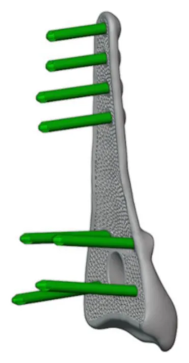
The customized medial malleolus implant incorporated specific design features to support anatomical reconstruction, fixation, and biological integration.
- Patient-Specific Geometry: Designed according to the patient’s anatomical requirements to reconstruct the missing medial malleolus.
- Porous Structure: A 3D Printed Ticellium porous structure from Jajal Medical, incorporated at the bone-contact surface to encourage bone ingrowth and osseointegration.
- Polished Surface Finish: The soft-tissue-facing surface was highly polished to minimize friction and potential soft-tissue irritation.
- Screw Fixation: Designed with multiple screw fixation points to provide stable implant positioning.
Patient-Specific Cutting Guide
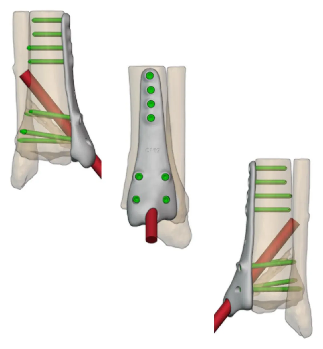
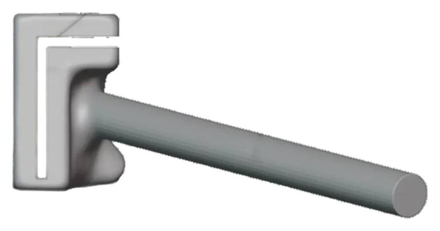
A patient-specific cutting guide was designed to facilitate accurate preparation of the affected medial malleolar region.
The guide was manufactured using medical-grade BioMed Amber material to support precise surgical positioning and resection planning.
This customized guide was developed to improve surgical accuracy and facilitate the placement of the patient-specific reconstruction implant.
Surgical Fixation
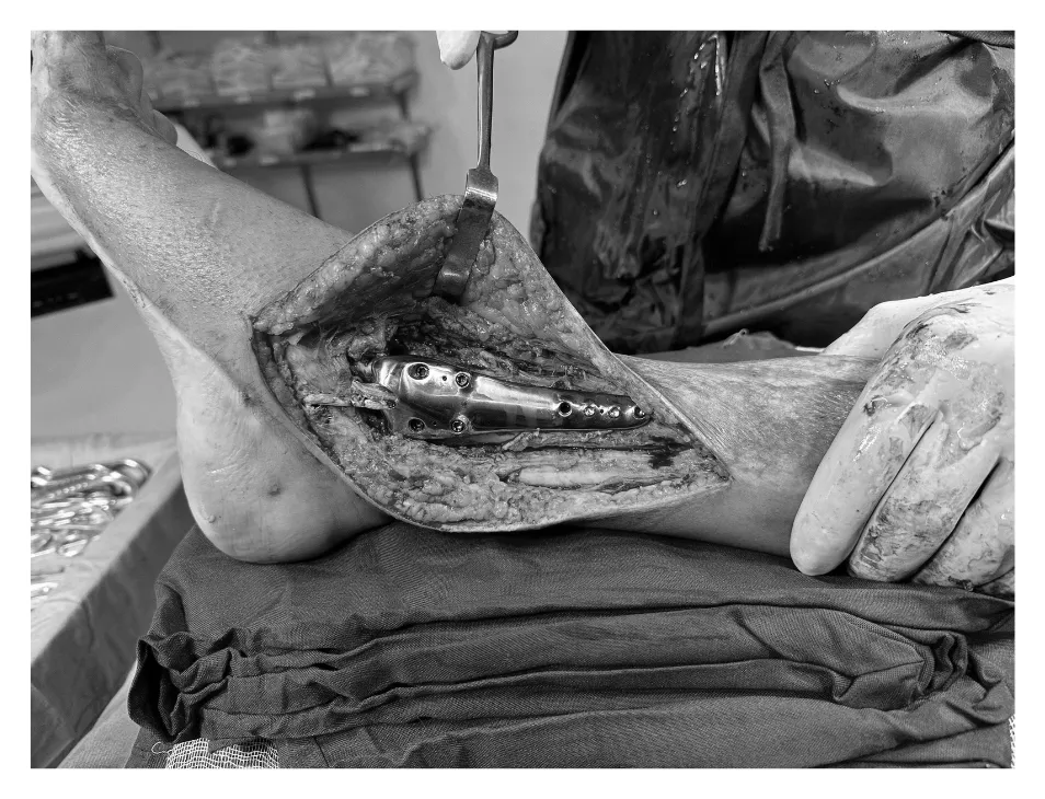
Following surgical preparation of the affected region, the customized Ti-6Al-4V medial malleolus implant was positioned and secured using 3.5 mm locking screws.
The implant’s customized geometry and fixation configuration were intended to restore the missing medial malleolar anatomy and provide structural stability to the ankle joint.
Clinical Outcomes
At the six-month follow-up, radiological evaluation demonstrated complete union with satisfactory implant positioning.
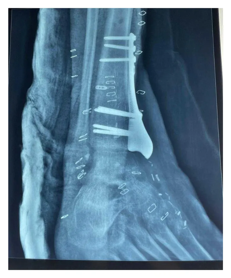
The patient achieved a functional ankle range of motion of approximately 45°, comprising 30° plantar flexion and 15° dorsiflexion.
Overall, the patient reported good satisfaction and successfully returned to unrestricted daily activities.
Related cases
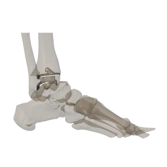 Ankle
Custom Total Ankle Replacement Using a Patient-Specific 3D-Printed Implant
A 48-year-old female with advanced osteoarthritis of the left ankle, where complex biomechanics and unique anatomy made conventional implant reconstruction difficult.
Read the case →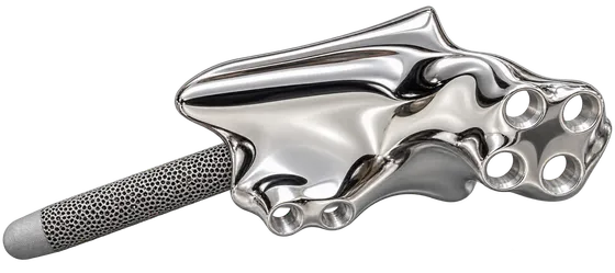 Tibia
Custom Tibia Implant for Complex Tibial Reconstruction
Mirrored from the healthy tibia to restore alignment and limb length
Read the case →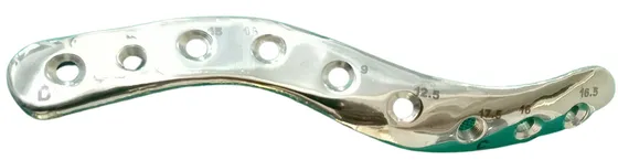 Clavicle
Patient-specific Clavicle Implant Plate
Clavicle malunion with brachial plexus compression treated by osteotomy and a mirrored custom plate
Read the case →Have a case like this?
Send the imaging and the clinical question — first 3D visualisation back within 24 hours.