
Club Foot Virtual Planning
Virtual osteotomy sequence and pre-op model for congenital talipes equinovarus
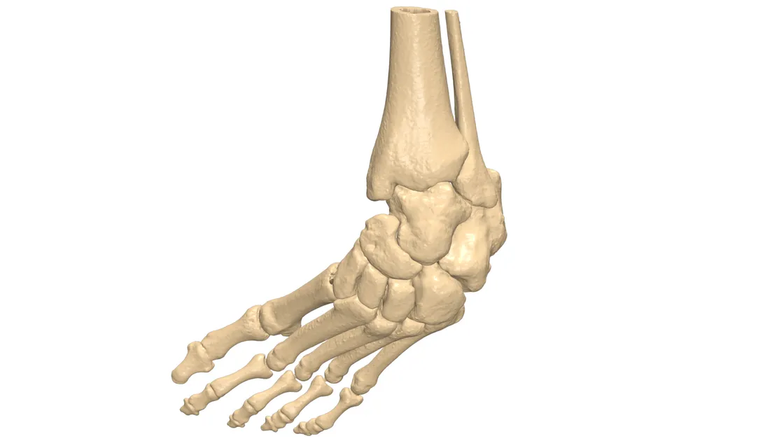
Case overview
Club foot is an infrequent deformity — a congenital foot condition in which the forefoot is turned inward and downward and cannot be straightened. It is also known as congenital talipes equinovarus.
A surgical approach can offer a radical form of treatment, with the goal of restoring the ankle joint. This case involved a 31-year-old male patient affected with club foot. With the surgical approach, better planning helps make surgery more accessible and accurate.
Jajal Medical played a vital role by virtually planning the surgery: mapping out the first visualisation, planning the procedure, and providing a 3D pre-op model for visualisation and better understanding.
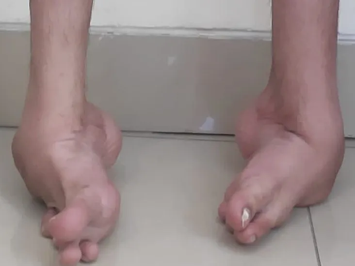
Segmentation
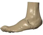
The region of interest was well captured by the scan and replicated in a 3D model for a better understanding of the deformity. The main goal was to provide a self-explanatory model helping the surgeon visualise information that can be missed in conventional 2D CT data.
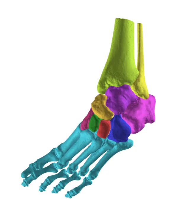
Virtual planning
3D visualisation and a pre-op model helped the surgeon understand and analyse the deformity and anatomy of the club foot.
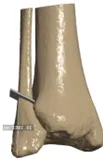
- The deformed talus bone was removed, then an osteotomy was performed on the distal part of the fibula
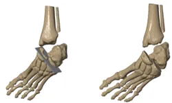
- Osteotomy was performed on the cuboid, navicular and calcaneus bones at the calcaneocuboid and talonavicular joints
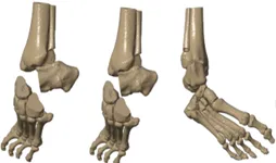
- The navicular and cuboid, along with all other bones, were positioned to the distal fibula at the calcaneus region
- External rotation with medial translation and advancement achieved the correct foot angle in line with the axis
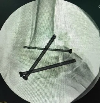
- The osteotomy was stabilised by fusion using fixation screws
Soft-tissue analysis
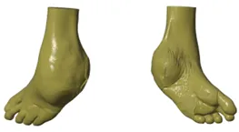
It was also necessary to understand the post-op soft-tissue alignment, and this was provided for better analysis.
Conclusion
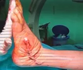
Pre-planning the surgical approach and using a 3D pre-op model helped achieve accurate surgical results, and a straight foot angle.
Related cases
 Ankle
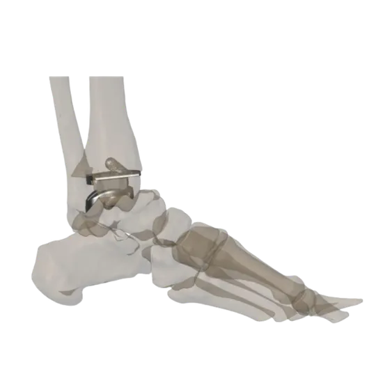
AnkleCustom Total Ankle Replacement Using a Patient-Specific 3D-Printed Implant
A 48-year-old female with advanced osteoarthritis of the left ankle, where complex biomechanics and unique anatomy made conventional implant reconstruction difficult.
Read the case → Oncology
Patient-Specific 3D Printed Hemipelvic Reconstruction with Constrained THA
Type I + II internal hemipelvectomy for Ewing's sarcoma
Read the case → Shoulder
Custom Hill-Sachs Implant for Reconstruction of a Large Engaging Humeral Head Lesion
Patient-specific implant and drilling guide for a 22 × 41 mm humeral head defect
Read the case →Have a case like this?
Send the imaging and the clinical question — first 3D visualisation back within 24 hours.