
Custom TMJ Implant
Total TMJ replacement and mandibular reconstruction after tumour removal, with a UHMWPE fossa
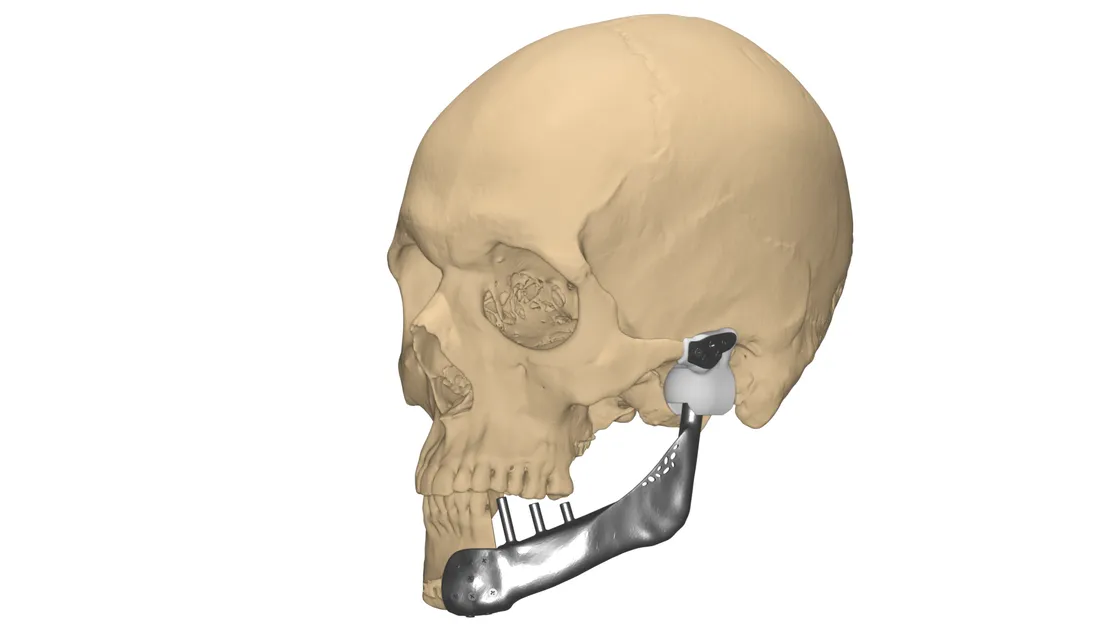
Case information
Temporomandibular joint (TMJ) disorder causes pain, stiffness or lack of mobility in the joint, preventing full range of jaw movement. Open joint surgery with replacement of the complete TMJ by an artificial prosthesis is required in severe TMJ disorder involving complex tumours or trauma defects.
A male patient aged 26 had undergone a primary tumour removal surgery in the past and required reconstruction of the deficient mandible with a customised implant. Dr. Vivek Saxena approached our clinical engineers for a robust plan and precise reconstruction of the TMJ using a patient-specific implant.
Visualisation
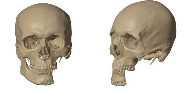
To visualise the mandibular anatomy and its neighbouring area, a high-resolution CT scan of adequate slice thickness and increment was used to 3D model the defect. The region of interest was replicated as a 1:1 digital model for understanding the defect and pathology — a self-explanatory model helping the surgeon visualise information that usually remains hidden in conventional 2D CT images.
Planning
Once segmentation was completed, the plan was to lay down the removal of the tumour followed by reconstruction using a customised TMJ.
- The tumour on the left mandible had been removed a year earlier including the condyle, so the right occlusion and articulation were essential when the TMJ was replaced
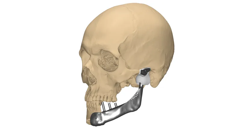
- The healthy right side was mirrored to reconstruct the naturally curved mandible and the osteotomy cut was decided
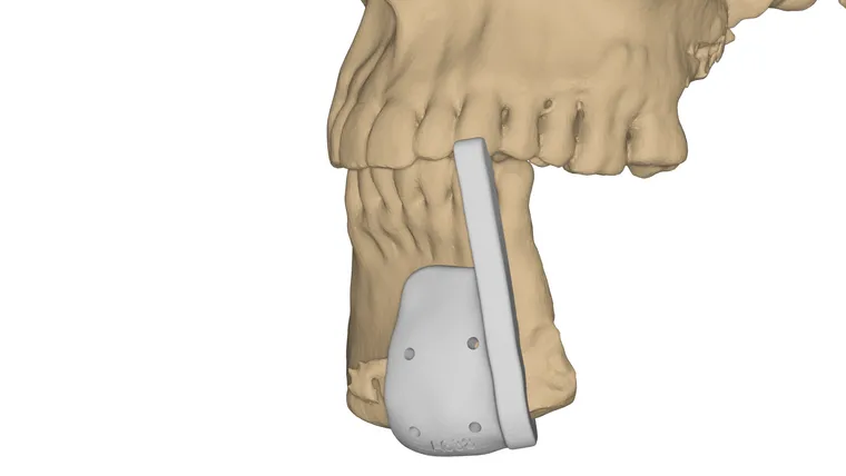
- A surgical guide was designed to resect the anterior mandible for a perfect joint connection of the implant body to the healthy right mandible
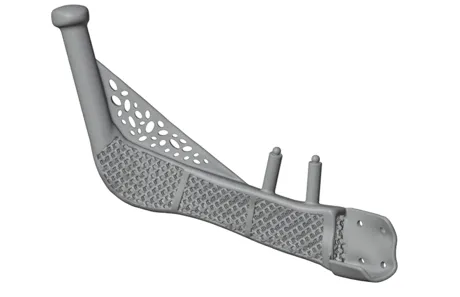
- A lattice structure inside the body reduced implant weight and aided osseointegration at the implant-bone contact surface
- Extruded dental abutment provisions were pre-defined in the mandible implant body at the desired positions to restore the teeth
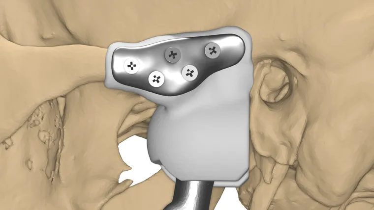
- Predefined holes — size, length and insertion trajectory based on bone density — on both body and fossa implant components
- The fossa was designed to ensure proper range of motion and natural jaw opening, preventing condylar dislocation
- A virtual simulation was carried out to ensure appropriate jaw movements with the implant in place
3D printing
Once the surgical plan was approved and finalised, it was translated into physical models to analyse the outcome. Pre-op, intermediate and post-op models were printed. The patient-specific guides were printed in biocompatible autoclavable material for realising the planned cuts intra-operatively on the mandible.
The customised implant was printed in medical-grade titanium, and the fossa was fabricated in high-strength medical-grade plastic — UHMWPE — with the required finishing and post-processing for better articulation.
Conclusion
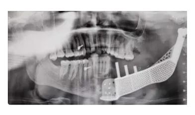
Pre-planning the surgical approach and using customised surgical guides and a patient-specific implant helped achieve accurate reconstruction, reduced intra-operative time and faster recovery of the patient.
This case was featured in the Times of India.
Related cases
 Jaw
JawCustomized Subperiosteal Implant
Peri-implantitis of maxilla and mandible in a 17-year-old, solved with custom titanium subperiosteal implants
Read the case →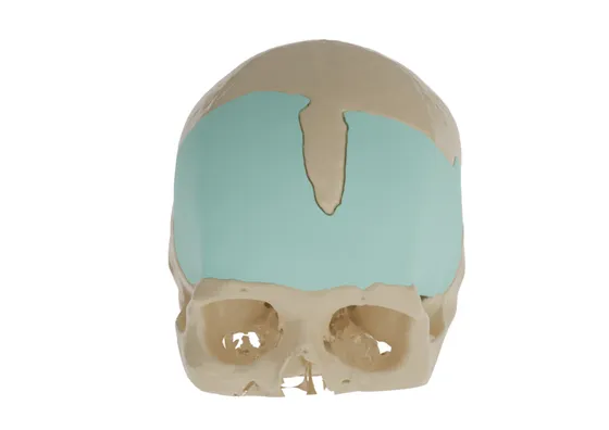 Cranial
Precision Reconstruction in Frontal Defect with Customized PMMA Cranial Implant
Two-part PMMA cranial plate restoring frontal bone curvature after a previous reconstruction
Read the case →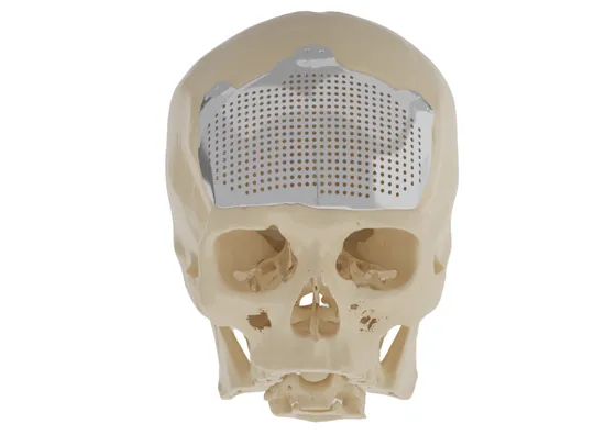 Cranial
Patient Specific Cranial Plate
Titanium cranioplasty designed to the healthy skull curvature after right-side skull involvement
Read the case →Have a case like this?
Send the imaging and the clinical question — first 3D visualisation back within 24 hours.