
Customized Subperiosteal Implant
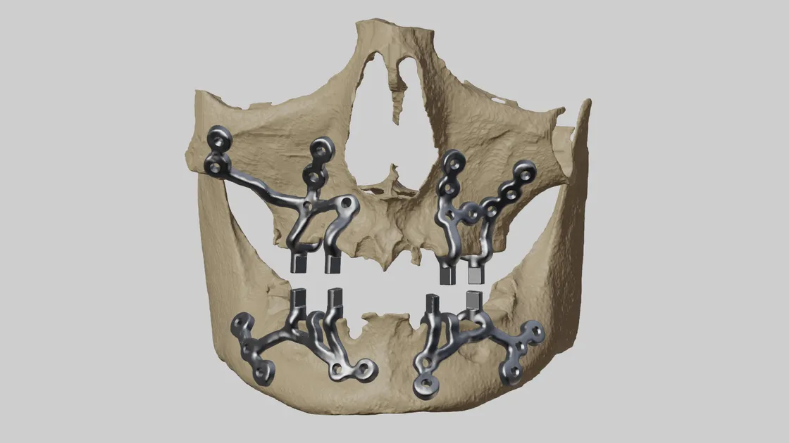
Peri-implantitis of maxilla and mandible in a 17-year-old, solved with custom titanium subperiosteal implants
Patient overview
A 17-year-old male presented with a severe case of peri-implantitis affecting both the maxilla and mandible regions. Due to loss of bone and extremely low density, a subperiosteal implant was the best option.
Challenges
- Thin bone structure: the patient’s thin bone posed a significant challenge for implant surgery — traditional methods were unsuitable due to inadequate bone density
- Maxilla and mandible deformity: peri-implantitis from previous implants in both regions required a comprehensive solution addressing both areas simultaneously
- Age and development: at only 17, the patient’s bone structure was still developing, requiring a tailored approach that would not interfere with ongoing growth
Solutions
Jajal Medical, in collaboration with Dr. Karthik R, developed a comprehensive surgical plan addressing the patient’s unique challenges.
- Advanced 3D planning software creating a precise surgical plan with detailed visualisation of the bone structure, enabling accurate implant placement and screw fixation
- Anatomical models providing a tangible representation of the surgical site, aiding planning and visualisation
- Proprietary software giving the surgical team a 3D view of the plan to analyse implant positions, angles and placement depth
- Medical-grade titanium chosen for biocompatibility and durability, ensuring the implants integrate with the bone over time
Procedure and outcome
Under the guidance of Dr. Karthik R, the surgery was performed using the advanced surgical plan. The implant surgery involved precise placement of subperiosteal implants in both the maxilla and mandible regions, with anatomical models and 3D visualisation facilitating accurate positioning and minimising complications.
The surgery was a resounding success. The patient’s thin bone structure was effectively addressed, and the implants were positioned optimally to support long-term stability and functionality.
Related cases
 Jaw
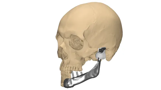
JawCustom TMJ Implant
Total TMJ replacement and mandibular reconstruction after tumour removal, with a UHMWPE fossa
Read the case →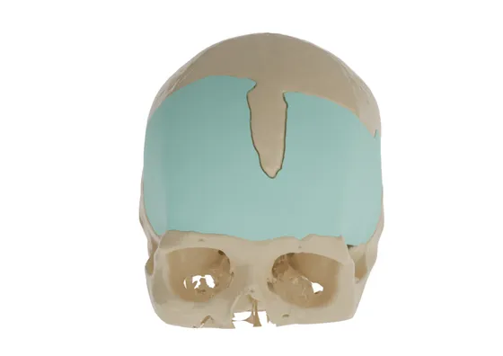 Cranial
Precision Reconstruction in Frontal Defect with Customized PMMA Cranial Implant
Two-part PMMA cranial plate restoring frontal bone curvature after a previous reconstruction
Read the case →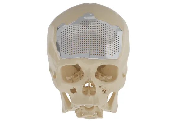 Cranial
Patient Specific Cranial Plate
Titanium cranioplasty designed to the healthy skull curvature after right-side skull involvement
Read the case →Have a case like this?
Send the imaging and the clinical question — first 3D visualisation back within 24 hours.