
Custom 3D-Printed Tibial Reconstruction for Pathological Fracture Secondary to Chronic Osteomyelitis
A two-stage hybrid reconstruction in a 12-year-old: infection control, then a custom TiCellium nail with a patient-specific PEKK graft
Patient overview
A 12-year-old female presented with pain over the left leg, inability to walk and intermittent fever.
She had previously been treated in Yemen for a suspected soft-tissue infection, undergoing three debridement procedures followed by intravenous antibiotic therapy at a local hospital. On examination, a healed surgical scar was noted with approximately 2 cm of shortening in the affected limb.
She was diagnosed with a pathological fracture of the tibial shaft secondary to chronic osteomyelitis. MRI and blood investigations were performed to assess the extent of infection and bone involvement.
Stage 1 — infection control and bone preparation
The first stage addressed the infection before any reconstruction could be considered:
- Extensive debridement and sequestrectomy to remove infected and non-viable bone.
- A cement spacer to manage the infected bone defect.
- Intraoperative samples taken for microbiological and culture analysis, with resected bone sent for histopathological examination.
- Discharge on culture-specific intravenous antibiotics with an above-knee slab.
- ESR and CRP monitored at regular intervals to assess the response to infection control.
Patient-specific surgical planning
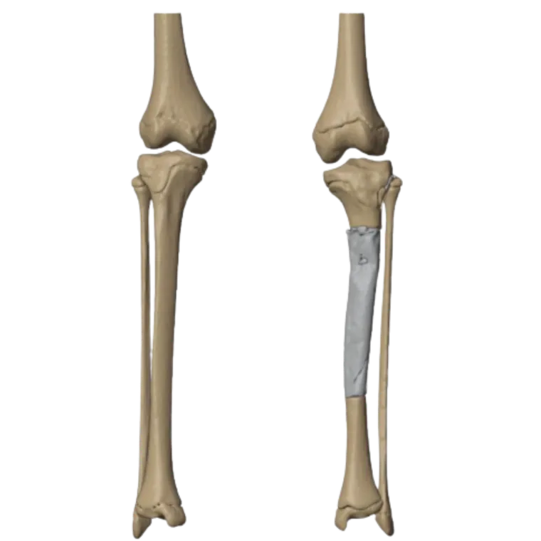
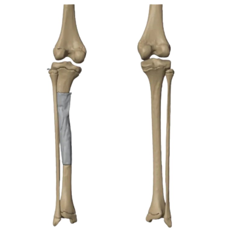
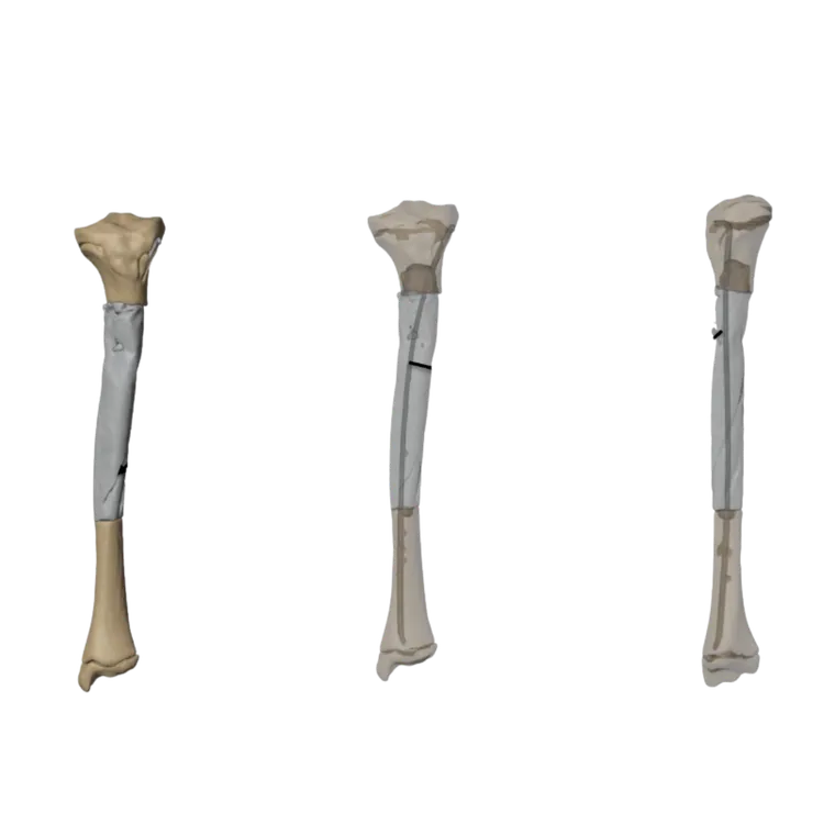
With the infection controlled, planning turned to the definitive reconstruction. A CT-based 3D assessment established the extent of bone loss and what the reconstruction would need to restore.
From the patient’s own anatomy and the segmental defect:
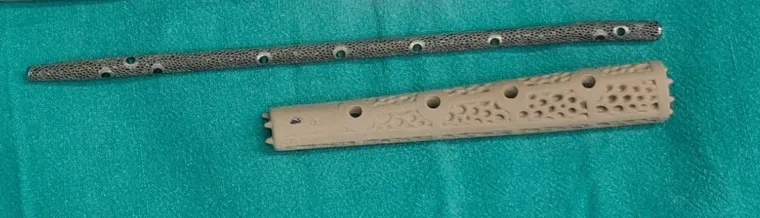
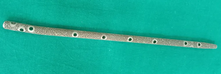
- A custom intramedullary nail was designed specifically for her, with measurements taken from the extent of bone loss and the anatomical requirements.
- A patient-specific PEKK graft was designed and manufactured by Oxford Performance Materials, USA.
- The components were planned together to bridge the tibial defect and provide structural support.
Stage 2 was scheduled once the customised implants were available.
Stage 2 — patient-specific tibial reconstruction
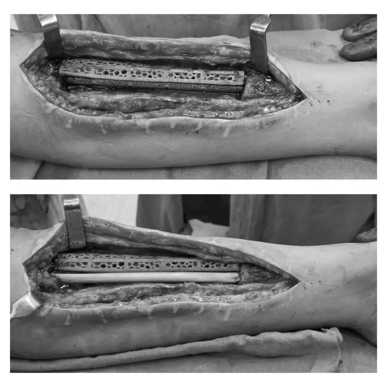
The definitive procedure combined spacer removal with fixation using the custom nail, the PEKK graft, autologous fibula grafting and soft-tissue reconstruction. The defect was bridged with:
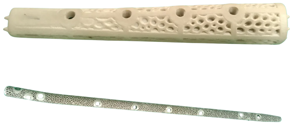
- A porous TiCellium 3D-printed custom nail
- The patient-specific PEKK implant
- An autologous fibula graft
- Locking fixation
- Soft-tissue reconstruction
Together these hybrid components were designed to bridge the defect, restore structural continuity of the tibia and provide stable fixation while incorporating biological bone grafting.
Custom 3D-printed reconstruction
The reconstruction was developed from the patient’s CT anatomy and the specific characteristics of the tibial defect.
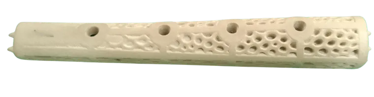
Key features: a custom nail matched to the patient’s anatomy and length; a porous TiCellium structure to support biological integration; a PEKK graft sized to the measured bone defect; an autologous fibula graft for biological reconstruction; locking fixation for mechanical stability; and integrated planning that addressed both the 2 cm shortening and the segmental bone loss.
Clinical impact
This case demonstrates the role of patient-specific 3D-printed implants and advanced digital planning in complex paediatric limb reconstruction following chronic osteomyelitis and pathological fracture.
Combining CT-based surgical planning, custom 3D-printed TiCellium technology, PEKK reconstruction, autologous fibula grafting and customised fixation produced a highly individualised hybrid strategy for a challenging tibial defect.
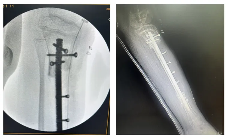
The case highlights how personalised implant design can support complex reconstruction where conventional options may be insufficient for the patient’s anatomy and the extent of bone loss.
Related cases
 Tibia
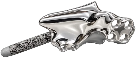
TibiaCustom Tibia Implant for Complex Tibial Reconstruction
Mirrored from the healthy tibia to restore alignment and limb length
Read the case →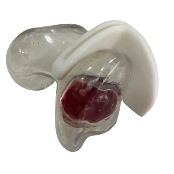 Femur
FemurPatient-Specific Cutting Jig for Distal Femur Tumor Resection
A CT-planned osteotomy guide for controlled tumour resection in the left distal femur
Read the case →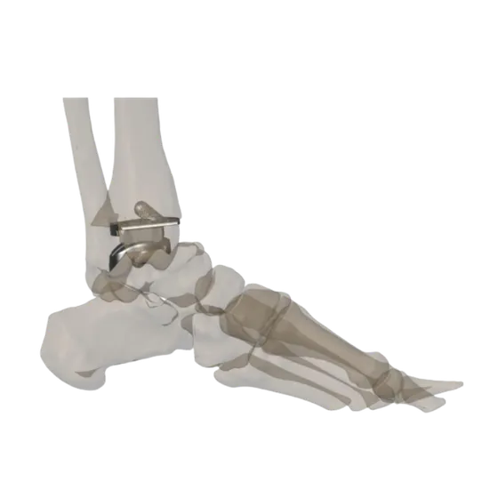 Ankle
AnkleCustom Total Ankle Replacement Using a Patient-Specific 3D-Printed Implant
A 48-year-old female with advanced osteoarthritis of the left ankle, where complex biomechanics and unique anatomy made conventional implant reconstruction difficult.
Read the case →Have a case like this?
Send the imaging and the clinical question — first 3D visualisation back within 24 hours.