
Patient-Specific Cutting Jig for Distal Femur Tumor Resection
A CT-planned osteotomy guide for controlled tumour resection in the left distal femur
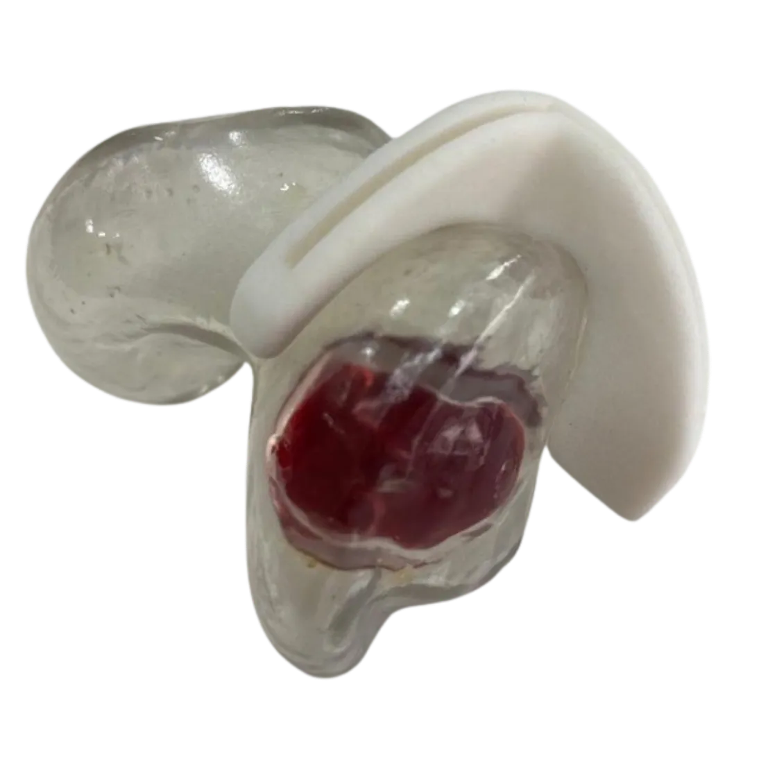
Patient overview
A 50-year-old male presented with significant pain and discomfort in the left knee, particularly during movement.
Preoperative CT imaging revealed the precise location, size and morphology of a tumour in the left distal femur. Because of the tumour’s position — and the need for accurate bone resection followed by reconstruction with a partial femoral implant — precise surgical planning was essential.
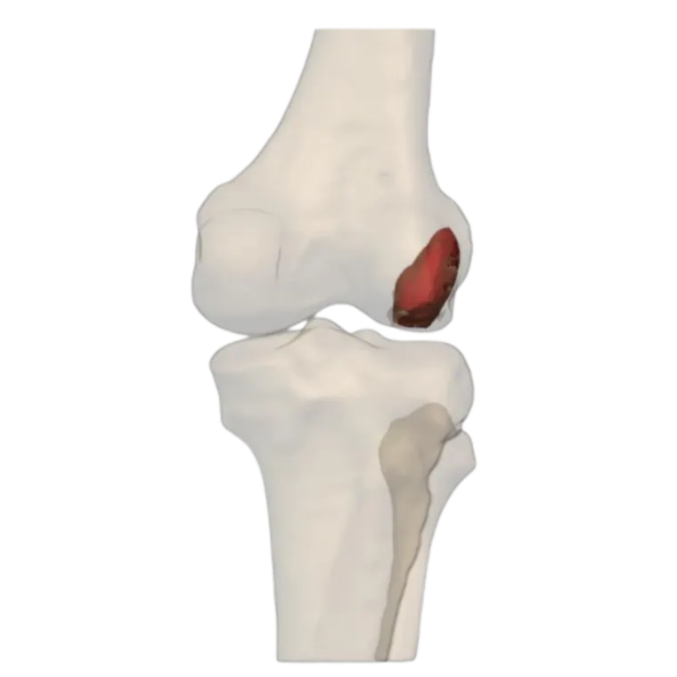
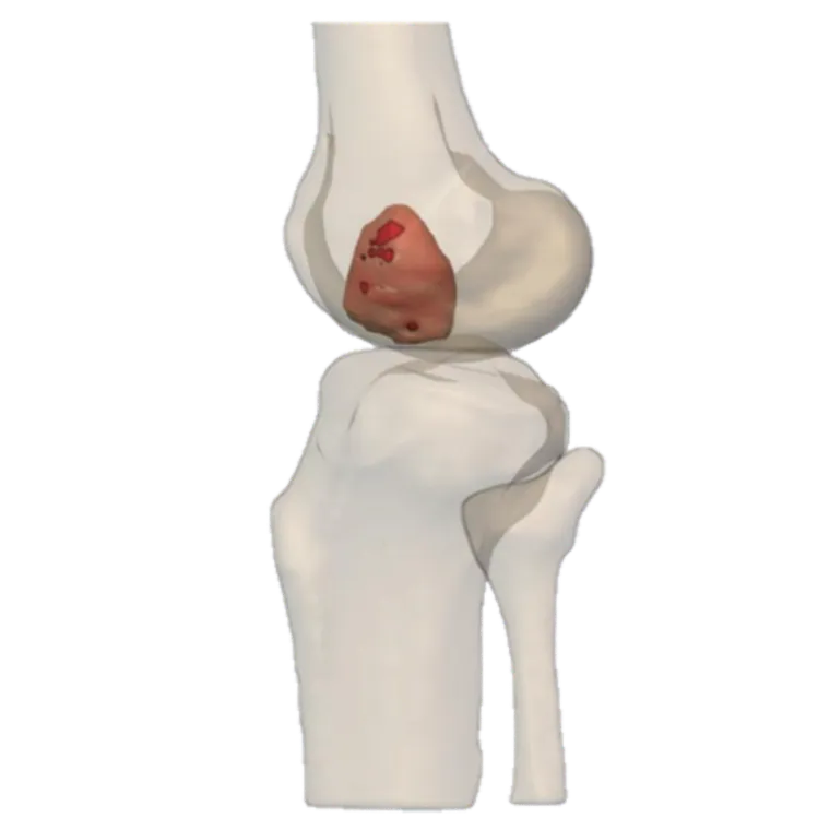
To facilitate accurate tumour removal, a patient-specific surgical osteotomy guide was designed around the patient’s individual anatomy and tumour characteristics.
Surgical planning
Working closely with the surgeon, the Jajal Medical design team developed a detailed patient-specific surgical plan.
- Preoperative CT imaging and 3D anatomical reconstruction were used to visualise the distal femur and pinpoint the tumour.
- The tumour boundaries were evaluated to determine the required osteotomy planes and resection margins.
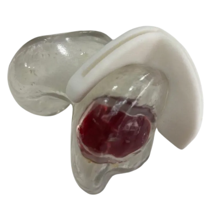
- A custom cutting jig was designed to conform precisely to the patient’s distal femoral anatomy.
- Guide geometry, cutting slots and fixation provisions were tailored to the planned surgical approach and intraoperative accessibility.
- The virtual surgical plan and guide design were reviewed to confirm positioning and execution before surgery.
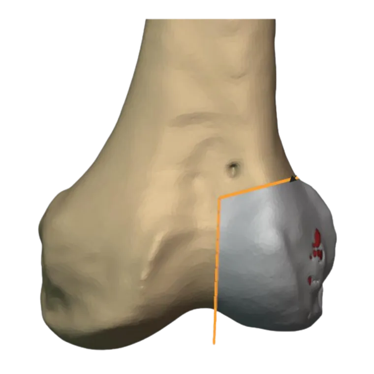
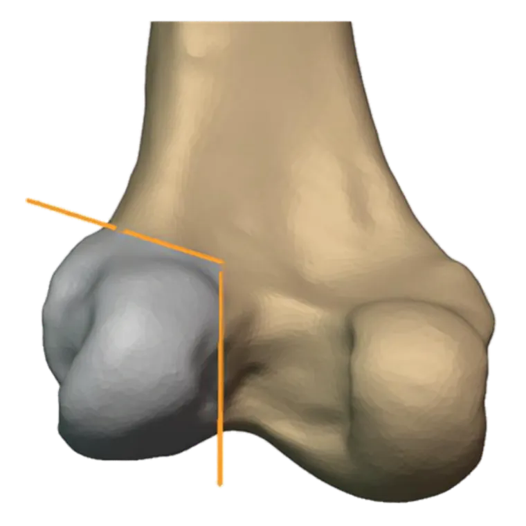
Patient-specific cutting jig
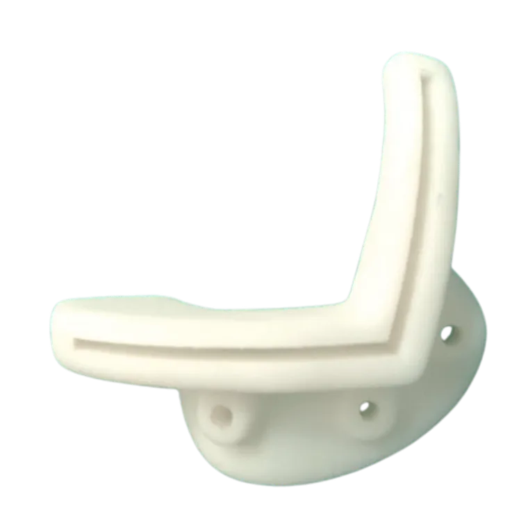
The custom cutting jig was developed to give the surgeon a precise and reproducible guide for tumour resection.
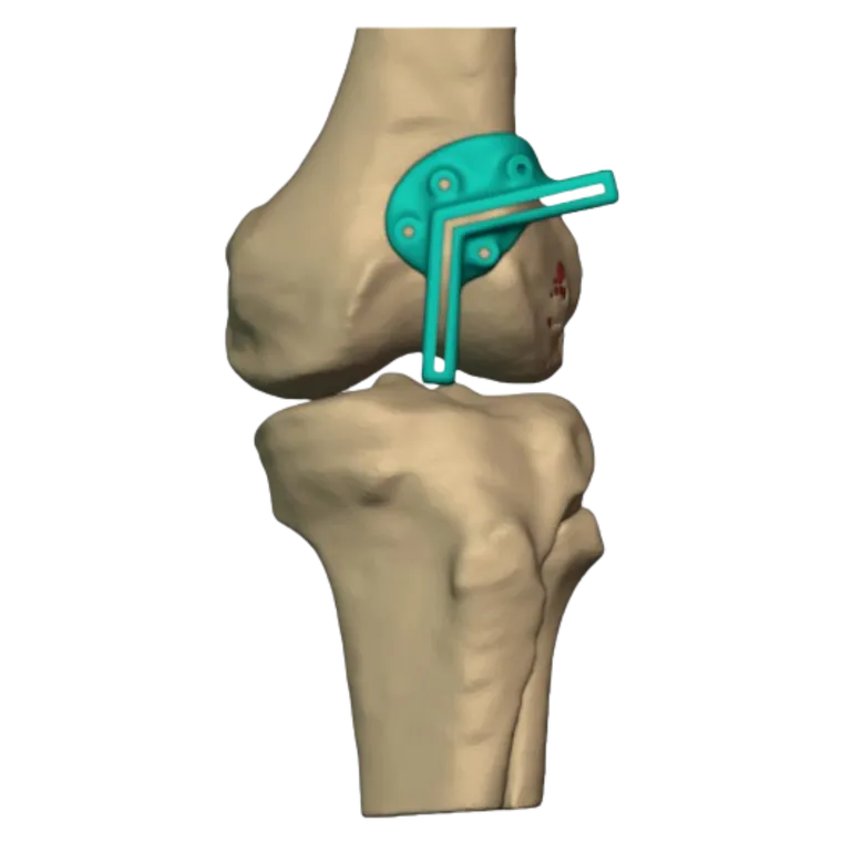
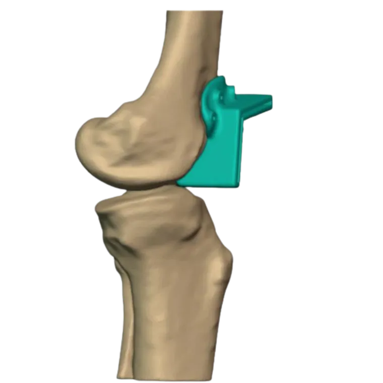
Key features of the design: patient-specific geometry matched to the distal femoral anatomy; cutting slots positioned to the planned osteotomy planes; dedicated fixation holes for stabilisation with K-wires; placement optimised for the surgical approach and intraoperative accessibility; and a form intended to support accurate resection and preparation for subsequent reconstruction.
Surgical procedure
During surgery the custom cutting jig was positioned onto the distal femur according to the preoperative plan.
- The guide was stabilised using K-wires through the dedicated fixation holes.
- Once securely positioned, the surgeon performed the osteotomy through the predefined cutting slots.
- The patient-specific guide helped maintain the planned cutting orientation and facilitated accurate removal of the tumour-bearing bone.
- Following resection, the femur was prepared for further reconstruction according to the surgeon’s requirements.
Clinical impact
This case demonstrates the value of patient-specific surgical guides in complex orthopaedic oncology procedures.
By combining CT-based 3D anatomical reconstruction, virtual surgical planning and customised guide design, Jajal Medical provided a solution tailored to the patient’s anatomy and tumour location.
The custom cutting jig supported accurate osteotomy, controlled tumour resection and efficient preparation for subsequent reconstruction — helping translate the preoperative surgical plan into the operating room with greater precision.
Related cases
 Femur
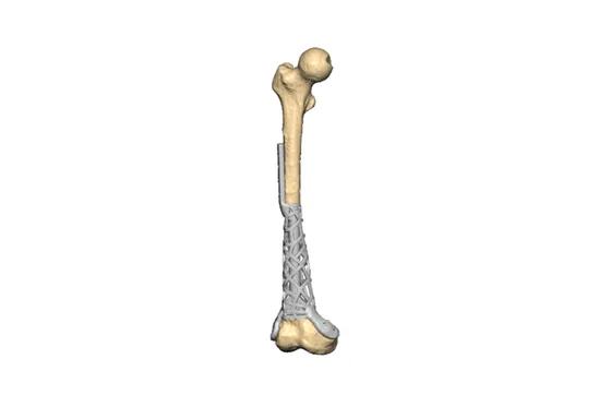
FemurDistal Femur Replacement
Custom intramedullary titanium implant after failed rod fixation, preserving the natural joint
Read the case →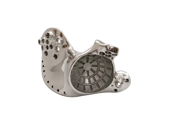 Oncology
Patient-Specific 3D Printed Hemipelvic Reconstruction with Constrained THA
Type I + II internal hemipelvectomy for Ewing's sarcoma
Read the case →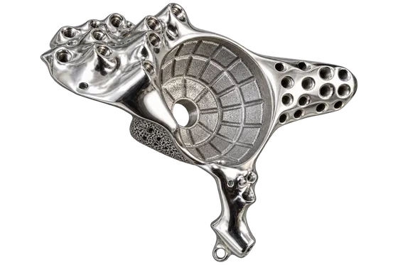 Pelvis
Pelvic Tumour Reconstruction Using Patient-Specific FTL Cup for Acetabular and Ischial Involvement
Planned resection, patient-specific jigs and custom fixation
Read the case →Have a case like this?
Send the imaging and the clinical question — first 3D visualisation back within 24 hours.