
Complex Hip Revision with Severe Acetabular Bone Loss Using Patient-Specific FTL Cup
Multi-flange fixation for a deficient acetabulum
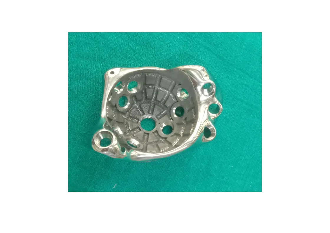
Patient overview
A 48-year-old female presented with a history of left-sided primary hip replacement. A subsequent revision had removed implants from the acetabulum and femur, after which bone cement was placed within the cavity.
The right hip joint was healthy; the left pelvis showed significant structural defects requiring reconstruction.
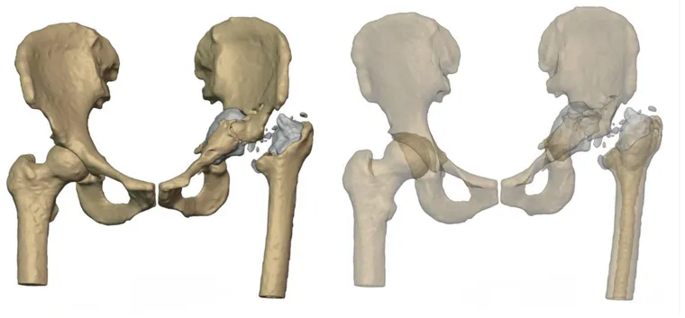
Preoperative assessment
CT imaging of the left pelvis revealed loss of the medial wall of the acetabulum, absence of the posterior column, a fracture in the anterior column, and an intact superior pubic ramus and ischium — a complex acetabular defect with compromised structural support.
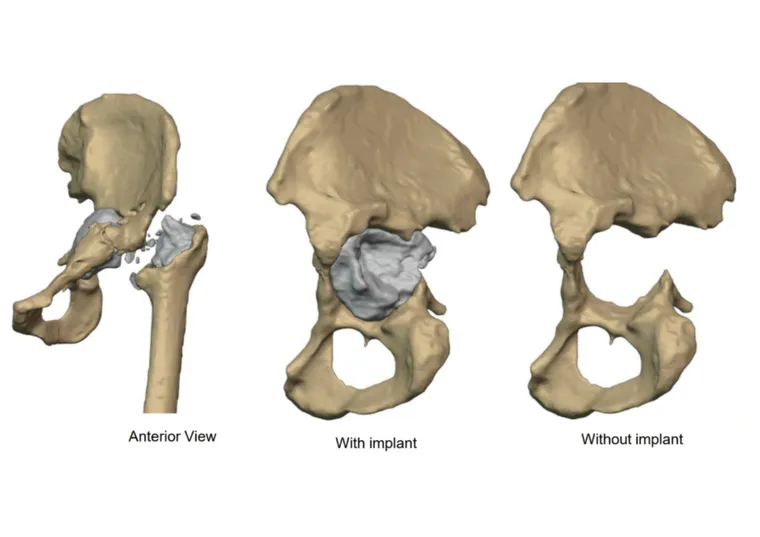
CT-based 3D surgical planning
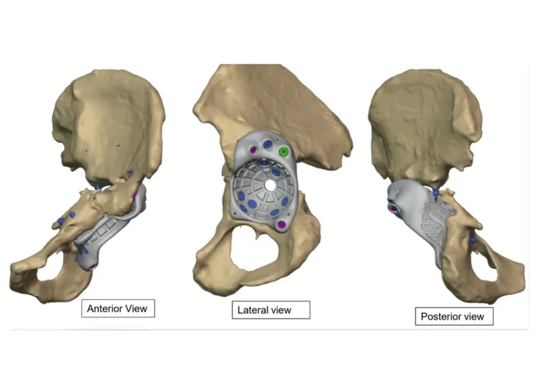
A CT-based 3D planning workflow was used to evaluate the defect and assess the available bone structure. On that basis the JMS team developed a patient-specific implant solution: a customised FTL (flanged triaxial locking) cup designed to cover the acetabular defect and facilitate load transfer.
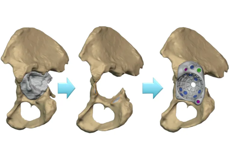
Key design features
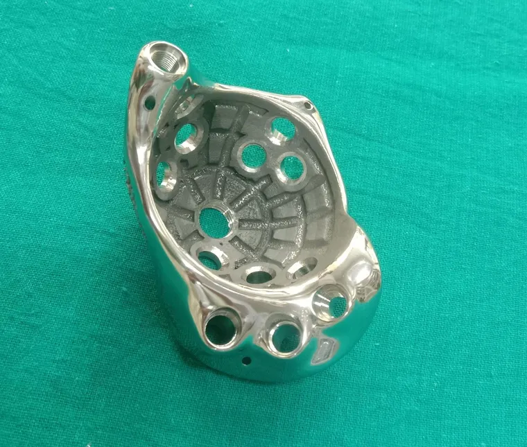
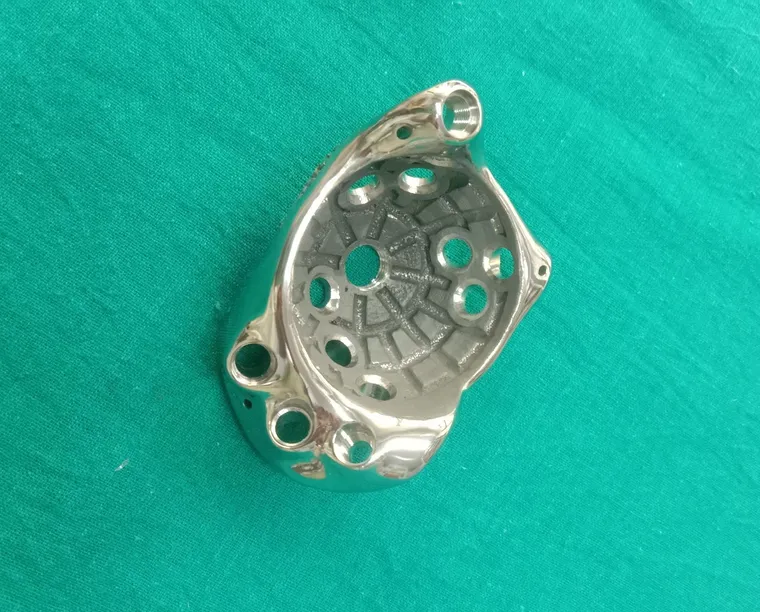
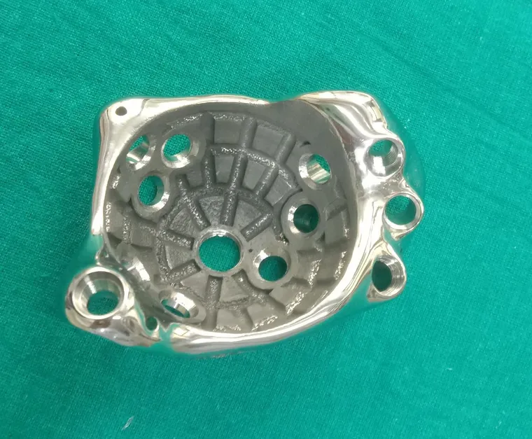
The FTL cup was designed to match the defect and cover the acetabular region. Three flanges extend to the pubis, ischium and ilium to support fitment and load transfer. Inclination and version angles were built into the design, clearance was provided for the surrounding soft tissues, and the cup diameter was defined to fill the defect while accommodating a standard cup and liner. The construct is secured with locking and compression screws.
Implantation strategy
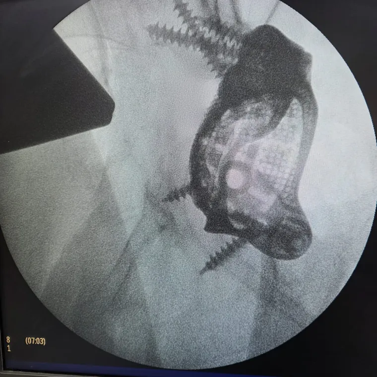
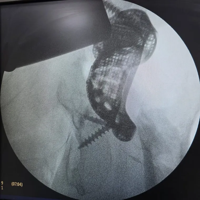
The FTL cup was positioned to cover the defect and achieve fixation against the pelvic bone, secured with locking and compression screws. Postoperative imaging confirmed coverage of the defect and implant fixation.
Clinical impact
This case demonstrates the application of a patient-specific FTL cup in managing a complex acetabular defect. CT-based planning and a customised implant enabled precise reconstruction in the presence of significant bone loss and structural deficiency.
Post-operative outcomes indicated stable implant positioning and enhanced functional recovery.
Related cases
 Revision hip
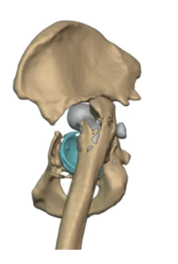
Customized FTL cup for Pelvic Discontinuity
Bilateral cup failure and pelvic discontinuity reconstructed with a custom FTL implant
Read the case →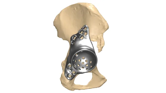 Revision hip
Custom Revision Hip Biflange Implant
Mirrored cage implant covering the acetabular defect after failed arthroplasty
Read the case →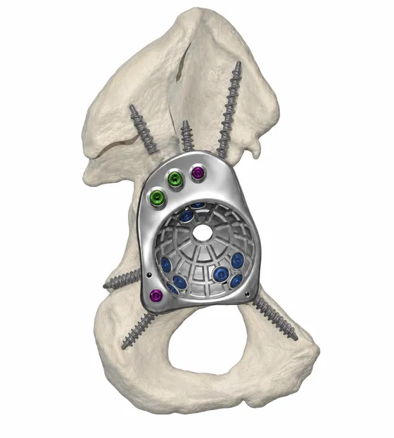 Hip
HipCustom FTL Cup for Complex Revision Hip Replacement
Massive anterior column bone loss reconstructed with a patient-specific FTL cup
Read the case →Have a case like this?
Send the imaging and the clinical question — first 3D visualisation back within 24 hours.