
Custom Revision Hip Biflange Implant
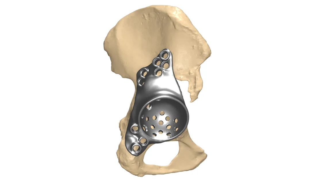
Mirrored cage implant covering the acetabular defect after failed arthroplasty
Case information
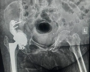
A 63-year-old female was having discomfort from a previous arthroplasty. The major complaint was pain and discomfort during mobility. The acetabulum and femur head were severely damaged.
The surgeon reached out to Jajal Medical for a customised implant. Based on her previous history and pre-op evaluations, a customised hip cup implant was chosen.
Visualisation
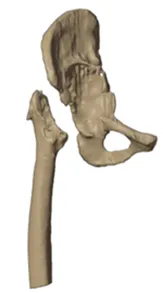
To visualise the hip anatomy and its neighbouring area, a high-resolution CT scan of adequate slice thickness and increment was used to 3D model the defect.
The region of interest was well captured in the scan and replicated as a 1:1 digital model for understanding the hip fracture. The idea is to provide a self-explanatory model helping surgeons visualise the defect that usually remains hidden in conventional 2D CT images.
Planning
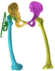
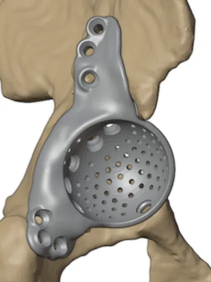
Based on the segmented data and 3D visualisation, we took the healthy side as reference and mirrored it to design the implant. Initial analysis was done on the deformed right side.
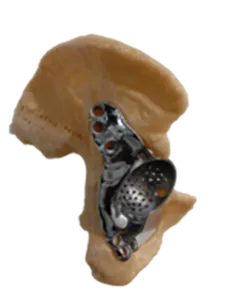
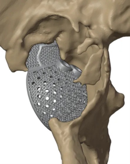
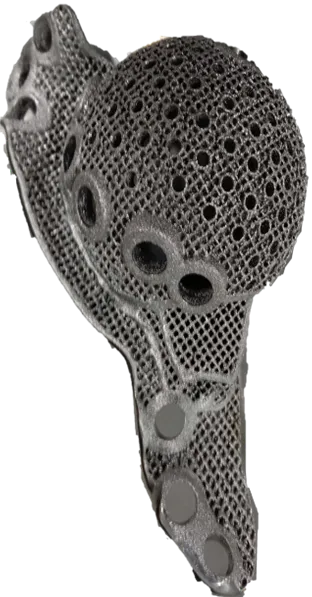
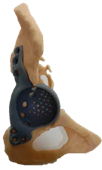
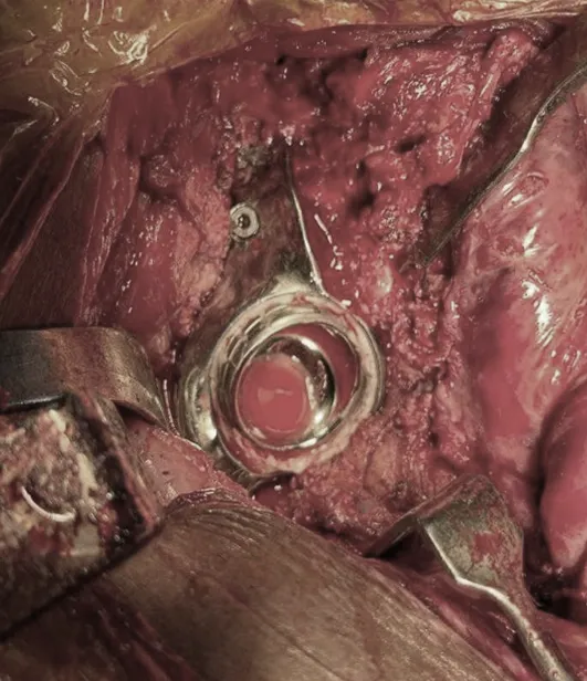
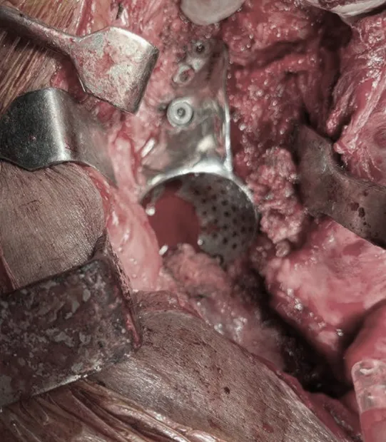
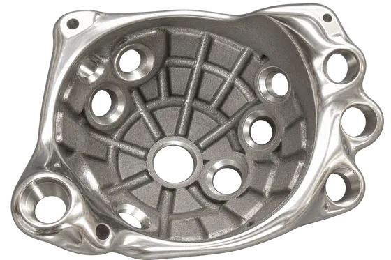
We reamed the bone and designed a customised cage implant covering the defect region on the surface of the hip, with the backside of the implant kept porous.
3D printing
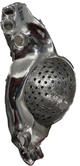
With the help of the design and planning, we 3D-printed a model of the deformed hip along with the implant, helping the surgeon visualise the pre-op condition and analyse the surgical approach against the expected post-op result.
Conclusion
Pre-planning the surgical approach and using the customised patient-specific implant helped achieve accurate reconstruction, reduced intra-operative time and faster recovery for the patient.
Related cases
 Revision hip
Complex Hip Revision with Severe Acetabular Bone Loss Using Patient-Specific FTL Cup
Multi-flange fixation for a deficient acetabulum
Read the case →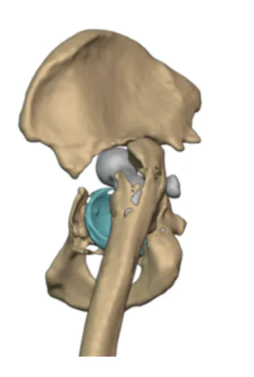 Revision hip
Customized FTL cup for Pelvic Discontinuity
Bilateral cup failure and pelvic discontinuity reconstructed with a custom FTL implant
Read the case →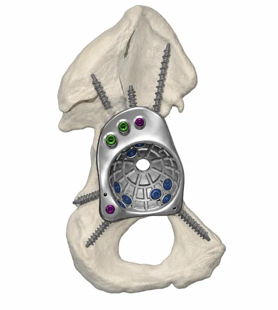 Hip
HipCustom FTL Cup for Complex Revision Hip Replacement
Massive anterior column bone loss reconstructed with a patient-specific FTL cup
Read the case →Have a case like this?
Send the imaging and the clinical question — first 3D visualisation back within 24 hours.