
Custom Hill-Sachs Implant for Reconstruction of a Large Engaging Humeral Head Lesion
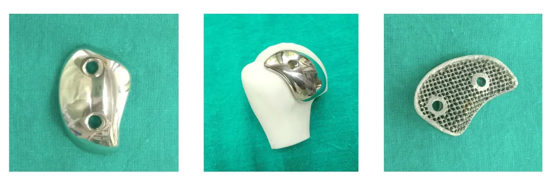
Patient-specific implant and drilling guide for a 22 × 41 mm humeral head defect
Patient overview
A 49-year-old female presented with recurrent anterior shoulder instability associated with a large Hill-Sachs lesion involving the left humeral head.
Preoperative imaging demonstrated an engaging humeral head defect that articulated with the anterior glenoid during shoulder motion, contributing to recurrent dislocation events. The defect measured approximately 22 mm × 41 mm, representing substantial bone loss and a significant compromise of the humeral articular surface.
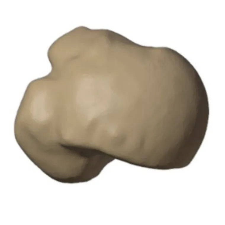
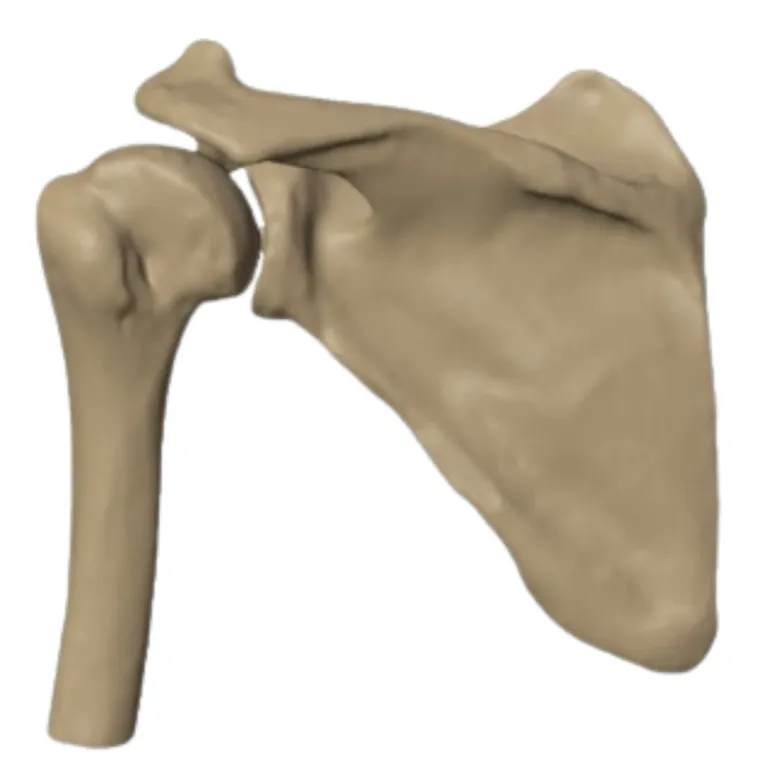
Surgical challenge
The dimensions and morphology of the lesion made the use of standard dome implants unsuitable for reconstruction.
Achieving anatomical restoration required an implant capable of matching the patient-specific geometry of the defect while allowing stable fixation within the available bone stock. As a result, a custom Hill-Sachs implant was developed to address the unique anatomical requirements of the case.
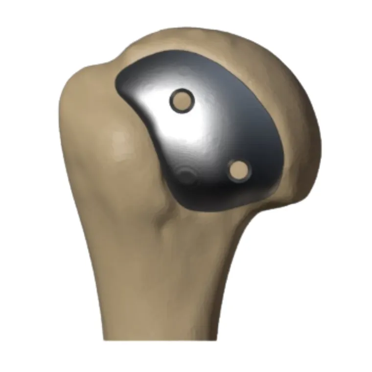
Patient-specific implant design
Using the patient’s imaging data, a custom implant was designed to accurately confirm the defect geometry and restore the native contour of the humeral head.
- Restore the articular surface anatomy
- Eliminate engagement between the humeral defect and the glenoid
- Re-establish humeral head congruity
- Provide stable mechanical fixation
Guided surgical execution
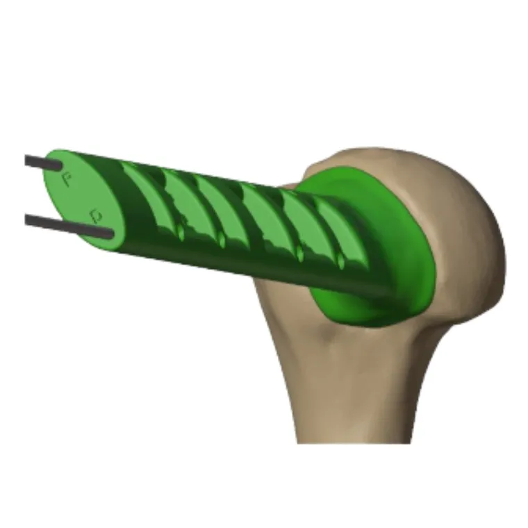
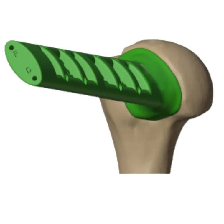
Following surgical exposure of the humeral head defect, a patient-specific drilling guide was positioned directly within the lesion.
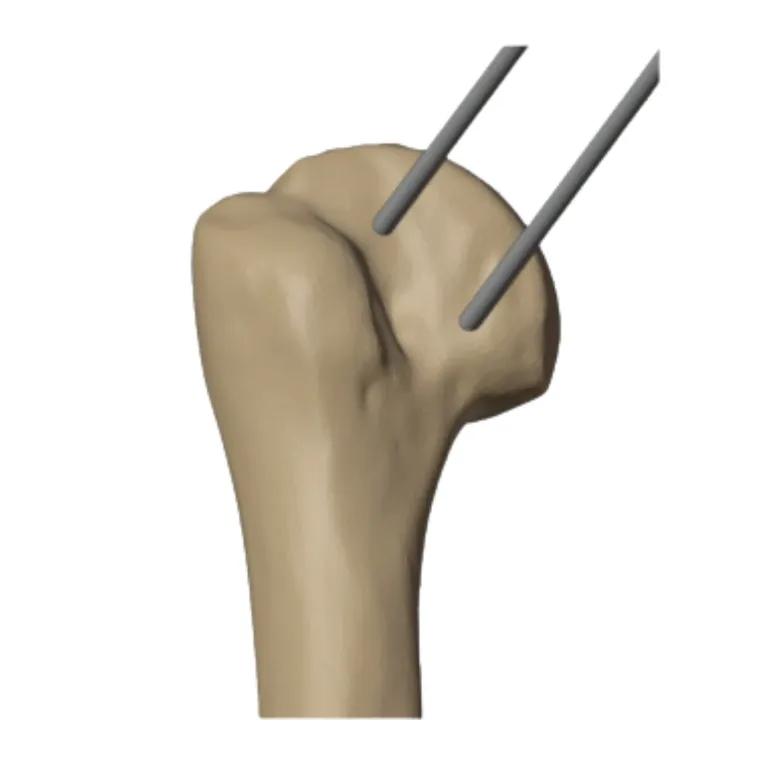
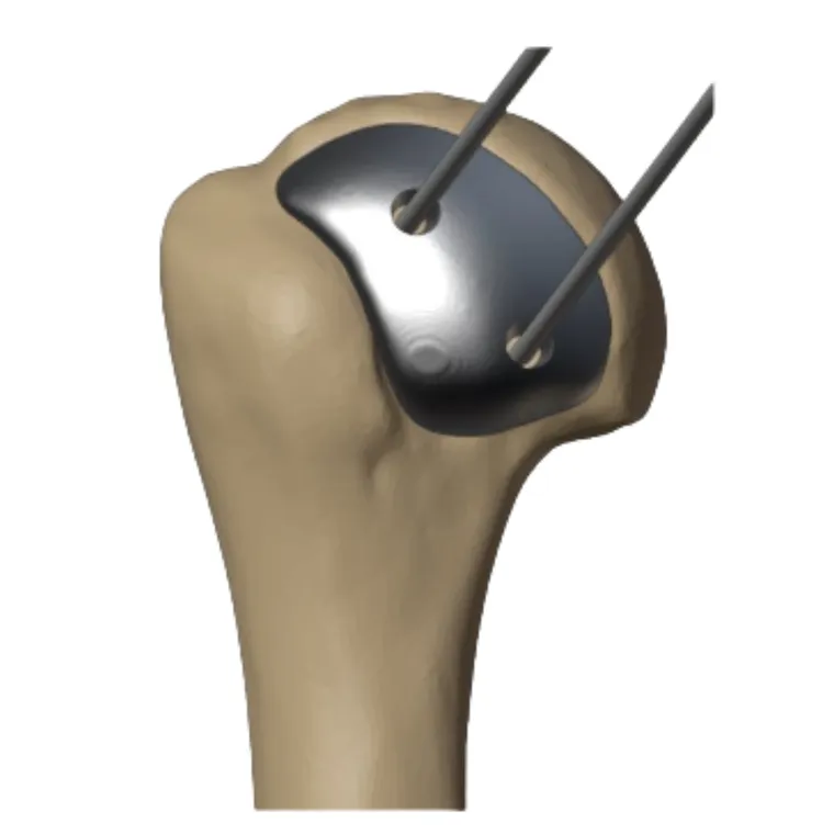
The guide facilitated the placement of two parallel K-wires, which provided the location and orientation for implant placement in the defect.
Post-op outcome
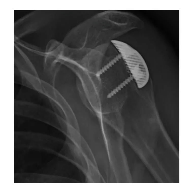
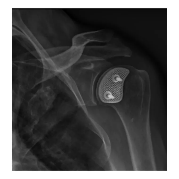
The reconstruction restored the humeral head contour and eliminated engagement with the glenoid, providing an anatomical solution designed to support joint stability and functional shoulder mechanics.
Clinical significance
Large engaging Hill-Sachs lesions remain a challenging source of recurrent shoulder instability, particularly when the extent of bone loss exceeds the limitations of conventional reconstructive options.
This case demonstrates the application of a patient-specific implant and instrumentation workflow for anatomical reconstruction of a substantial humeral head defect. The integration of preoperative planning, patient-specific guides, guided implant placement and stable fixation enabled a structured approach to restoring humeral head anatomy and addressing instability where standard implant solutions were not feasible.
Related cases
 Shoulder
Reverse Shoulder Arthroplasty with a Patient-Specific Custom Glenoid Implant
ReGlen custom base plate for severe glenoid bone loss in rheumatoid disease
Read the case → Shoulder
Custom ReGlen Implant for Complex Glenoid Reconstruction
Revision after failed shoulder arthroplasty with severe glenoid bone loss
Read the case →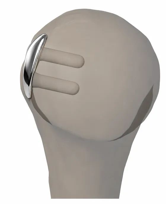 Shoulder
World's First Custom Implant for Hill Sachs Lesion
A pioneering “Fill Sachs” implant engineered to the patient's own humeral head curvature
Read the case →Have a case like this?
Send the imaging and the clinical question — first 3D visualisation back within 24 hours.