
Custom 3D-Printed FTL Cup for Complex Acetabular Reconstruction Through the Direct Anterior Approach
Extensive acetabular bone loss with posterior column destruction, reconstructed with a patient-specific 3D-printed titanium FTL cup and drilling guide
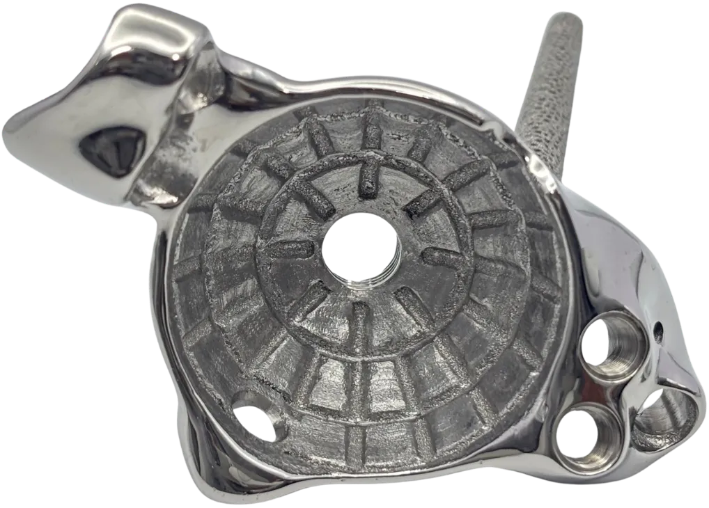
- Patient: 64-year-old female with multiple myeloma
- Problem: Extensive acetabular bone loss, posterior column destruction and posterior dislocation of the femoral head; unable to walk
- Solution: Patient-specific 3D-printed titanium FTL (Flanged Triaxial Locking) acetabular cup with a patient-specific bone drilling guide
- Fixation: Sciatic buttress peg, iliac compression and locking screws, pubic screw and two ischial screws
- Approach: Entirely through the Direct Anterior Approach (DAA) with a tensor fasciae latae (TFL) snip
- Result: Postoperative radiographs showed a well-seated implant construct, satisfactory fixation and restoration of the hip centre
Patient overview
A 64-year-old female patient with multiple myeloma presented with extensive acetabular bone loss, destruction of the posterior column of the acetabulum and posterior dislocation of the femoral head, which left her unable to walk.
Because the acetabular defect was so severe and lacked adequate structural support, conventional off-the-shelf implants were considered insufficient to give the stability required. A patient-specific 3D-printed titanium FTL acetabular implant was therefore designed for the complex anatomy and to support reconstruction through the Direct Anterior Approach (DAA).
Clinical challenge
The acetabulum was severely compromised. The posterior column was extensively destroyed and structural support had been lost. The posteriorly displaced femoral head made the reconstruction more complicated.
The primary surgical challenges were:
- Addressing extensive acetabular bone deficiency
- Achieving stable implant fixation despite posterior column destruction
- Restoring the anatomical hip centre
- Establishing fixation across the available pelvic bone
- Performing the entire reconstruction through the Direct Anterior Approach
A customised implant was needed to fit the patient’s unique anatomy and give a planned fixation strategy.
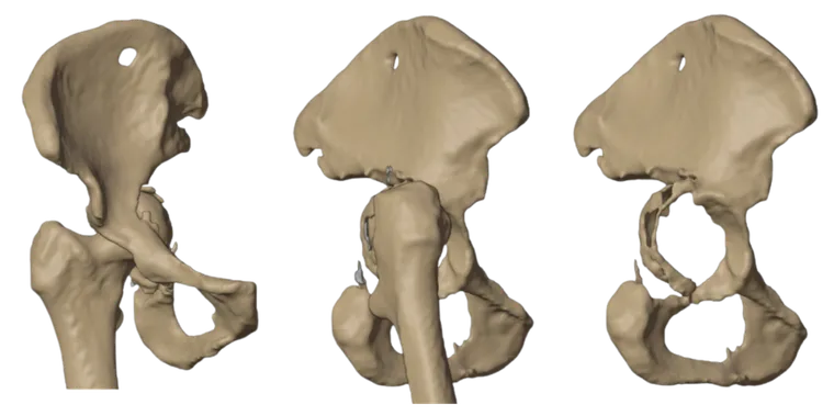
Patient-specific FTL cup design
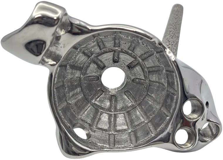
A custom 3D-printed titanium FTL cup was developed from CT-based preoperative planning and tailored to the patient’s acetabular defect.
Porous lattice structure (3D Printed Ticellium porous structure from Jajal Medical)
A porous implant surface was incorporated to support bone ingrowth and biological fixation.
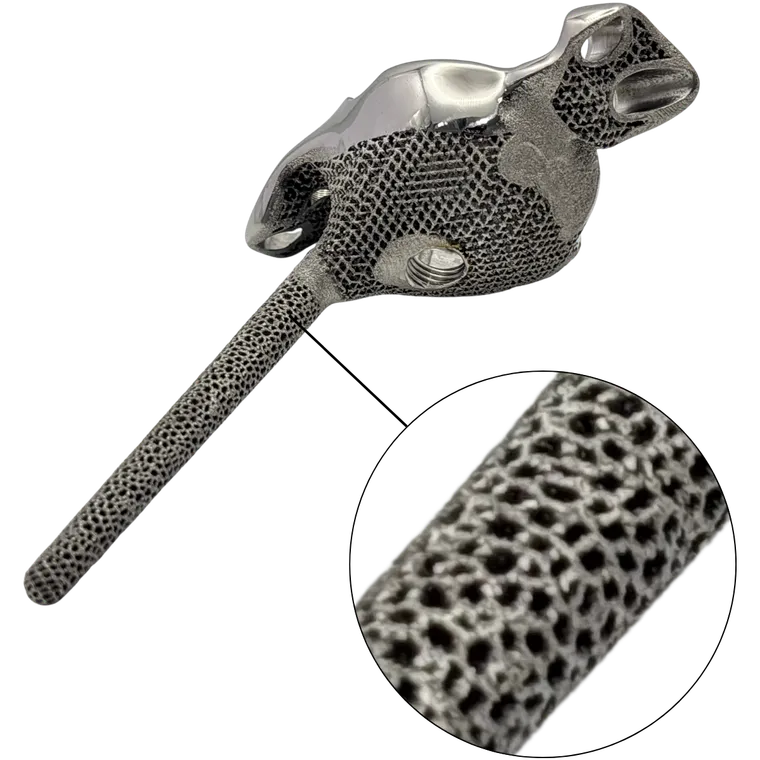
Sciatic buttress fixation
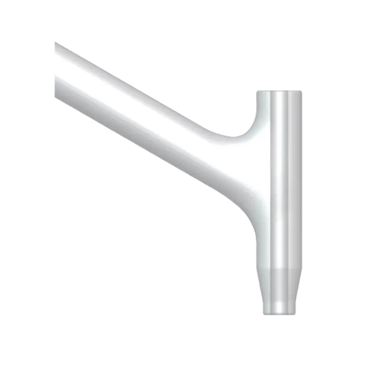
A dedicated buttress peg was directed towards the sciatic buttress. It gives additional support where the posterior column is no longer adequate.
Iliac fixation
Planned iliac fixation with compression and locking screws was incorporated to increase construct stability.
Inferior hemisphere fixation
Pubic screw fixation and two ischial screws were incorporated to give additional fixation across the inferior pelvis.
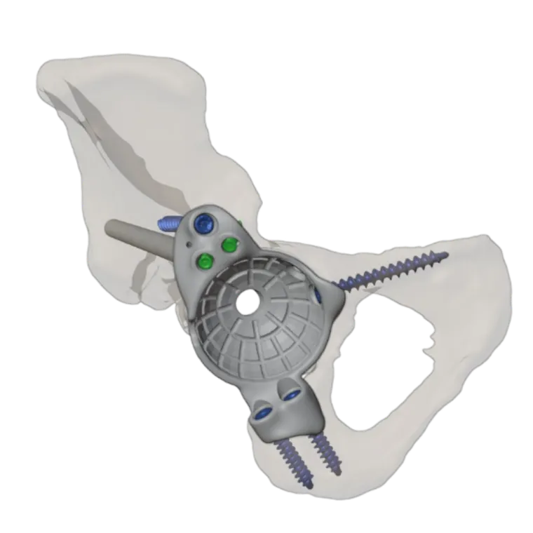
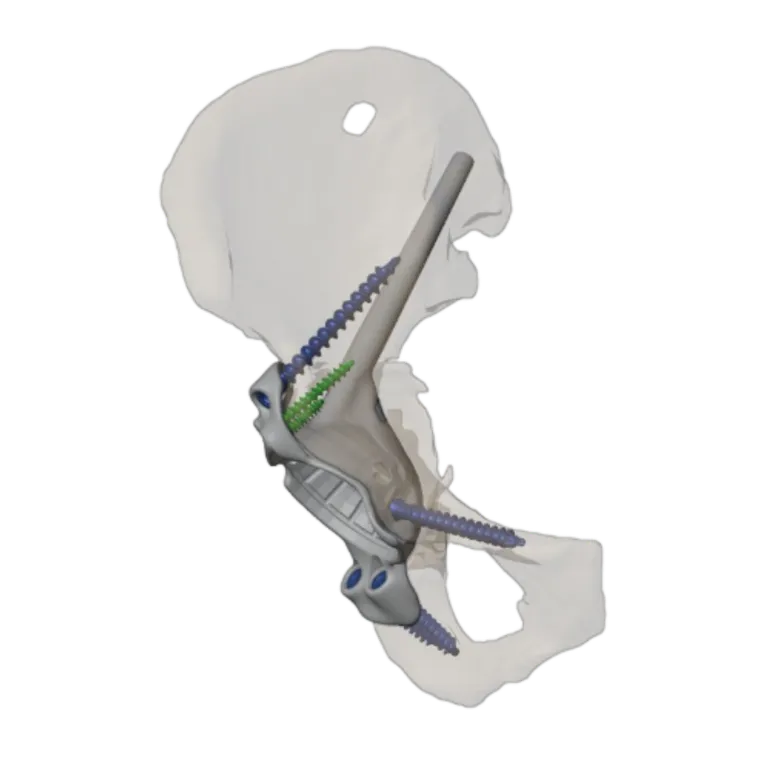
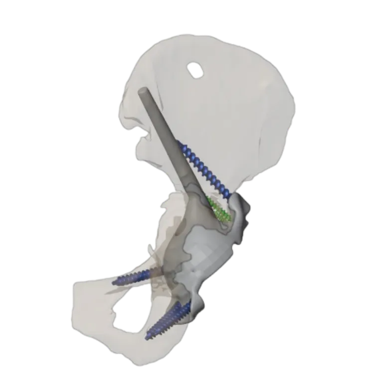
The patient-specific design aimed to optimise implant positioning, make use of the available bone stock and establish a stable foundation for the acetabular reconstruction.
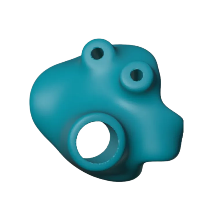
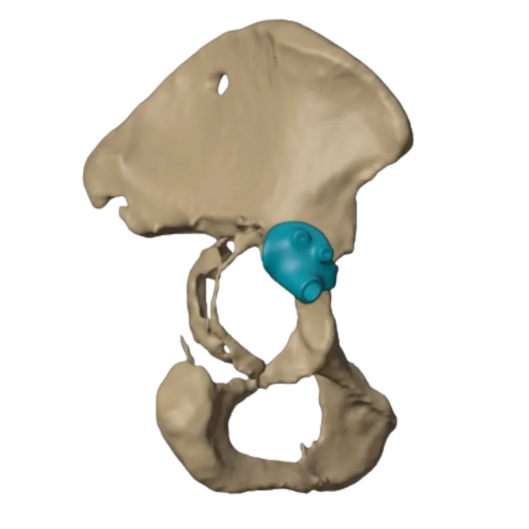
Patient-specific drilling guide for peg and screw fixation
A patient-specific bone drilling guide was designed from the CT-based preoperative plan to help with accurate positioning and drilling for the planned peg and screw fixation. The guide supported the intended trajectories for the dedicated sciatic buttress peg and for the iliac, pubic and ischial fixation screws.
This customised instrumentation was meant to carry the virtual surgical plan into the operating room. It helps achieve precise implant placement and optimise fixation in the presence of extensive acetabular bone loss and compromised posterior column support.
Surgical approach: reconstruction through the Direct Anterior Approach
The acetabular reconstruction was performed entirely through the Direct Anterior Approach (DAA), with a tensor fasciae latae (TFL) snip to give the required exposure.
This approach let the surgical team carry out the planned reconstruction with the customised FTL implant. The procedure involved positioning the patient-specific implant and following the planned fixation strategy, including iliac, sciatic buttress, pubic and ischial fixation.
The customised implant and CT-based planning were used to address the complex defect while restoring the hip centre and achieving stable fixation.
Post-op outcome
Postoperative radiographs showed a well-seated implant construct, satisfactory fixation and restoration of the hip centre.
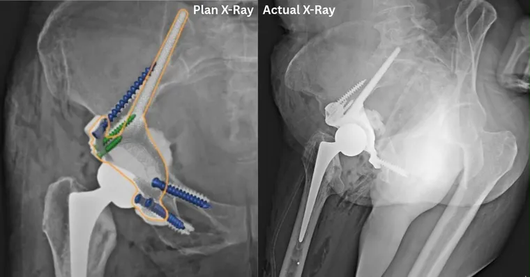
These findings supported the technical objectives of the reconstruction. Long-term implant survivorship, functional recovery and clinical outcomes need follow-up assessment.
Clinical impact
Custom FTL cup implants are valuable reconstructive options in selected cases of severe acetabular bone loss, where conventional implants may not give adequate fixation.
This case shows the potential role of patient-specific 3D-printed implants, CT-based surgical planning and a tailored fixation strategy in managing complex acetabular defects.
The reconstruction was performed entirely through the Direct Anterior Approach, which is an uncommon approach for this technically demanding procedure. To the best of the treating surgeon’s knowledge, this may be the first reported case in India of custom FTL acetabular reconstruction for such an extensive defect performed entirely through the Direct Anterior Approach.
Frequently asked questions
What is an FTL cup?
The FTL (Flanged Triaxial Locking) Cup is Jajal Medical's custom, patient-specific acetabular component. It is designed from the patient's CT scan for severe acetabular bone loss. In this case, fixation was planned into the ilium, the sciatic buttress, the pubis and the ischium.
Why was a custom implant used instead of an off-the-shelf cup?
The acetabular defect was so severe and lacked so much structural support that conventional off-the-shelf implants were considered insufficient to give the required stability.
How was fixation achieved without an intact posterior column?
A dedicated buttress peg was directed towards the sciatic buttress. It was combined with iliac compression and locking screws, a pubic screw and two ischial screws.
Which surgical approach was used?
The reconstruction was performed entirely through the Direct Anterior Approach (DAA), with a tensor fasciae latae (TFL) snip for exposure.
How was the virtual plan transferred to surgery?
A patient-specific bone drilling guide, designed from the CT-based plan, guided the trajectories for the sciatic buttress peg and the iliac, pubic and ischial screws.
Related cases
 Hip
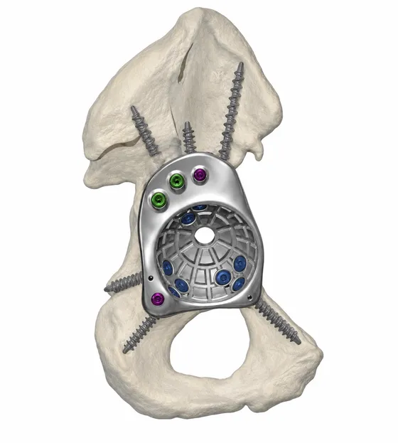
Custom FTL Cup for Complex Revision Hip Replacement
Massive anterior column bone loss reconstructed with a patient-specific FTL cup
Read the case →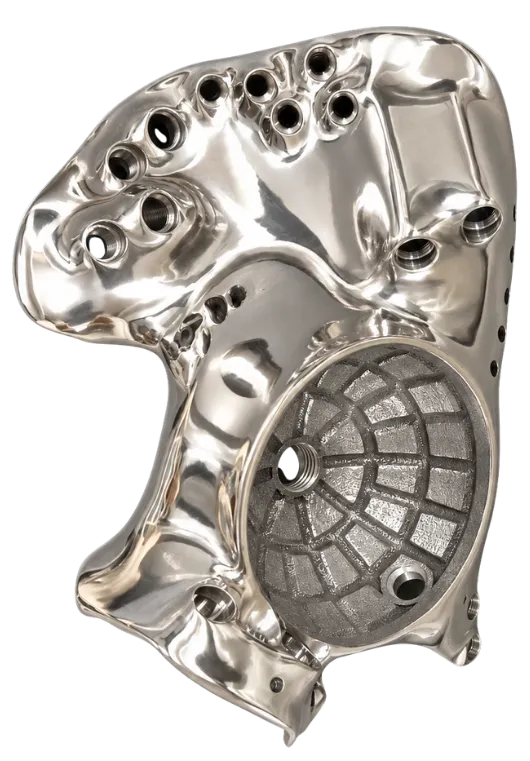 Oncology
Patient-specific FTL Cup for Tumor Resection and Acetabular Reconstruction
Pelvic tumour resection with cutting guides and a custom FTL cup
Read the case →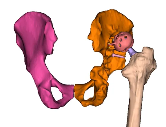 Hip
Complex Hip Revision
Ruptured medial wall and vertical hardware shift, segmented through heavy metal artifact
Read the case →Have a case like this?
Send the imaging and the clinical question — first 3D visualisation back within 24 hours.