
Patient-Specific 3D Printed Hemipelvic Reconstruction with Constrained THA
Type I + II internal hemipelvectomy for Ewing's sarcoma
Patient overview
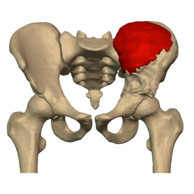
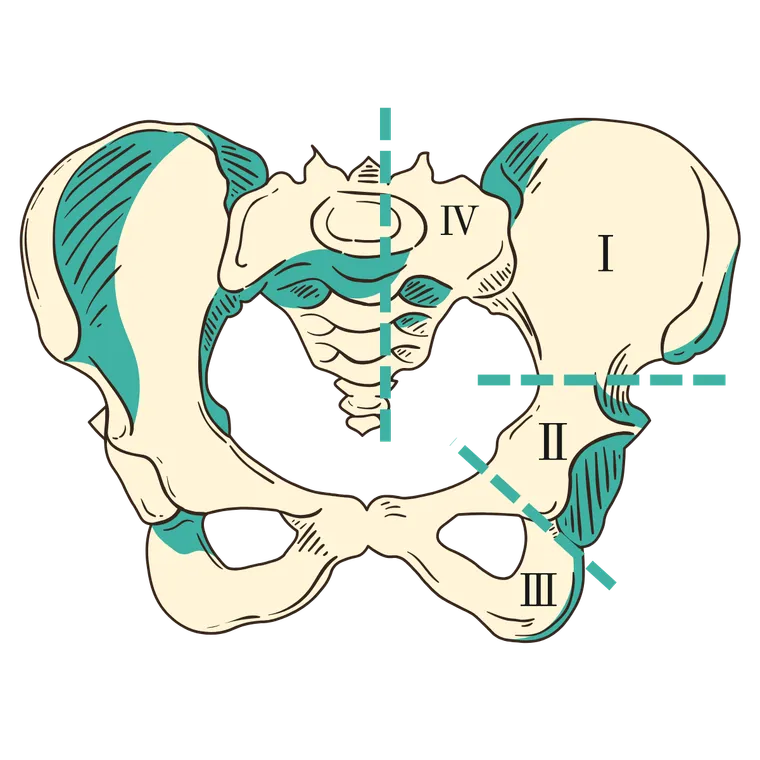
A 20-year-old male with metastatic Ewing’s sarcoma of the left hemipelvis presented following induction chemotherapy. Imaging revealed extensive involvement of the iliac wing and acetabulum, requiring a Type I + II internal hemipelvectomy.
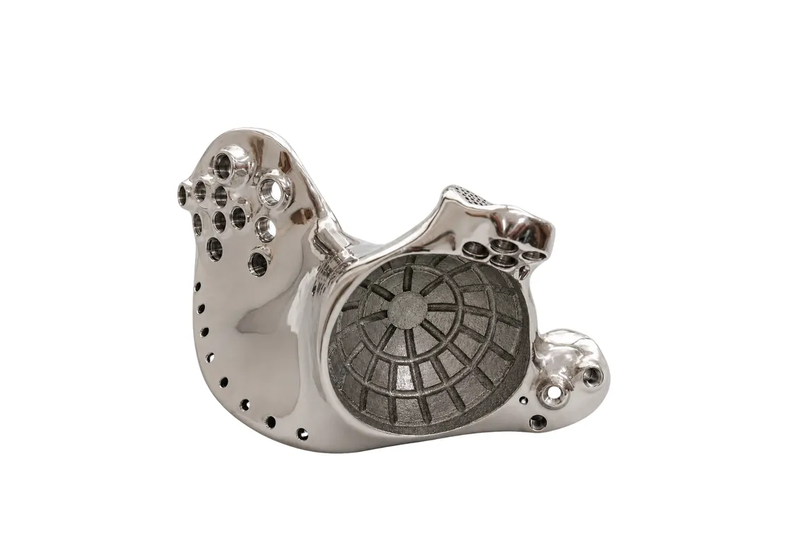
Given the complexity of the defect and the patient’s young age, a patient-specific 3D printed hemipelvic implant was designed to restore pelvic anatomy, preserve limb function, and provide a stable foundation for hip reconstruction.
The clinical challenge
Reconstruction after periacetabular tumour resection remains one of the most demanding procedures in orthopaedic oncology. Conventional techniques — biological grafts and standard pelvic prostheses — bring implant instability, limited fixation due to bone loss, infection risk, limb-length discrepancy, delayed rehabilitation and variable functional outcomes.
For this patient the goal was complete tumour resection while restoring pelvic continuity and enabling an early return to mobility.
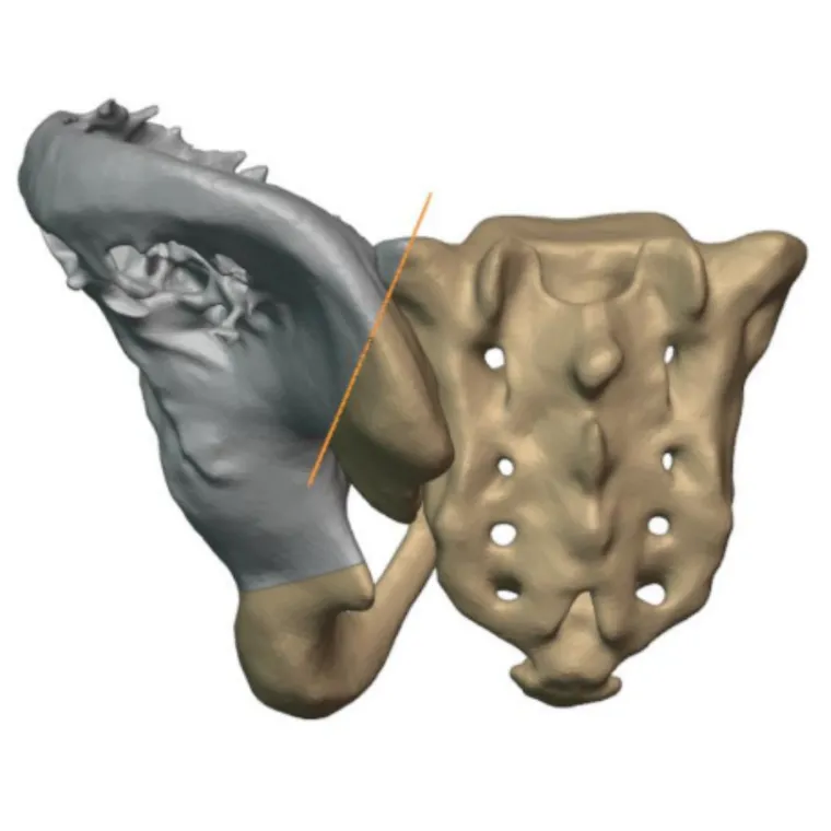
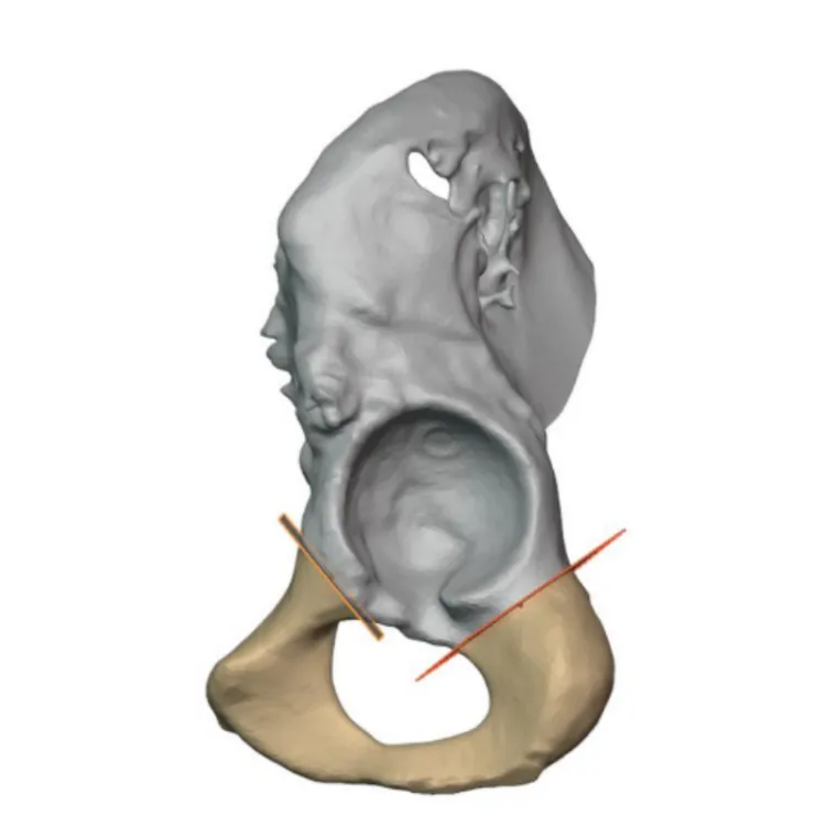
Personalised surgical planning
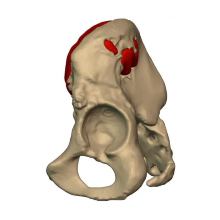
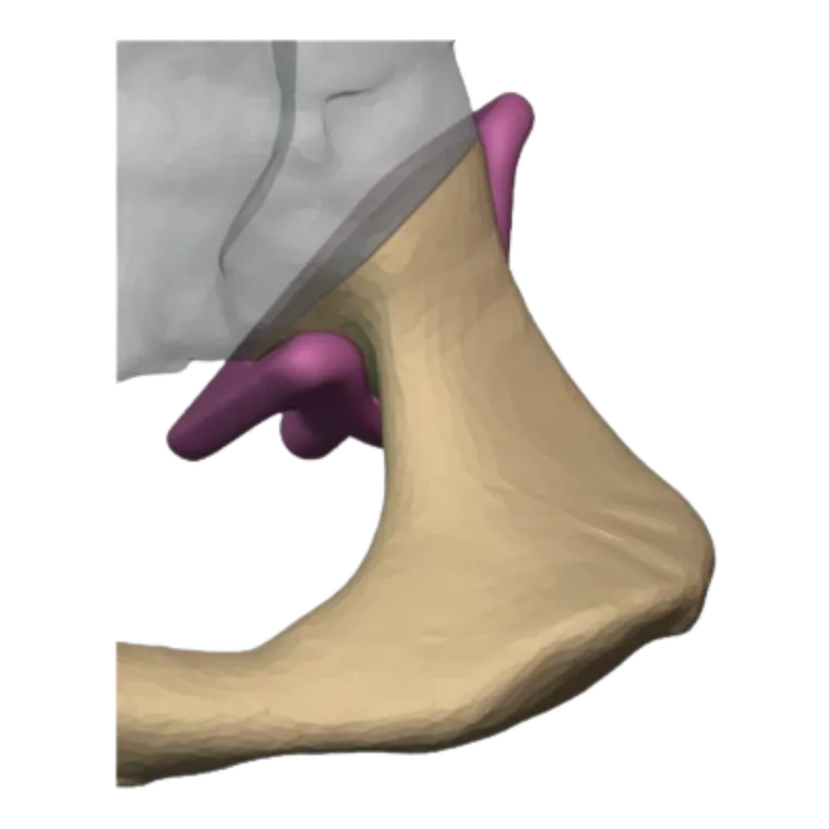
A CT-based three-dimensional model of the pelvis enabled comprehensive preoperative planning. The digital workflow produced resection planes, patient-specific resection guides for precise osteotomies, a customised porous titanium hemipelvic implant, optimised iliosacral fixation and a constrained total hip arthroplasty construct.
The implant was engineered to restore native hip biomechanics while maximising primary stability and long-term biological fixation.
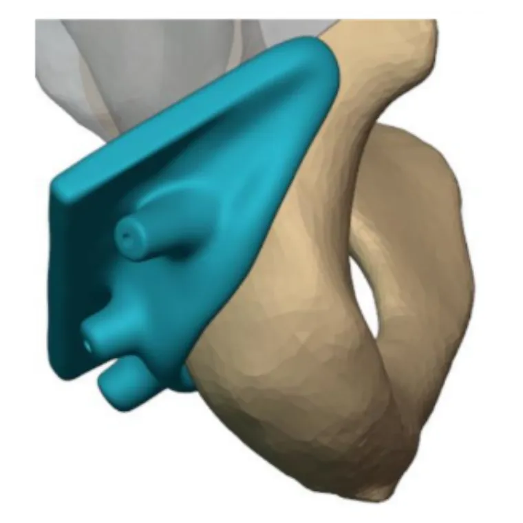
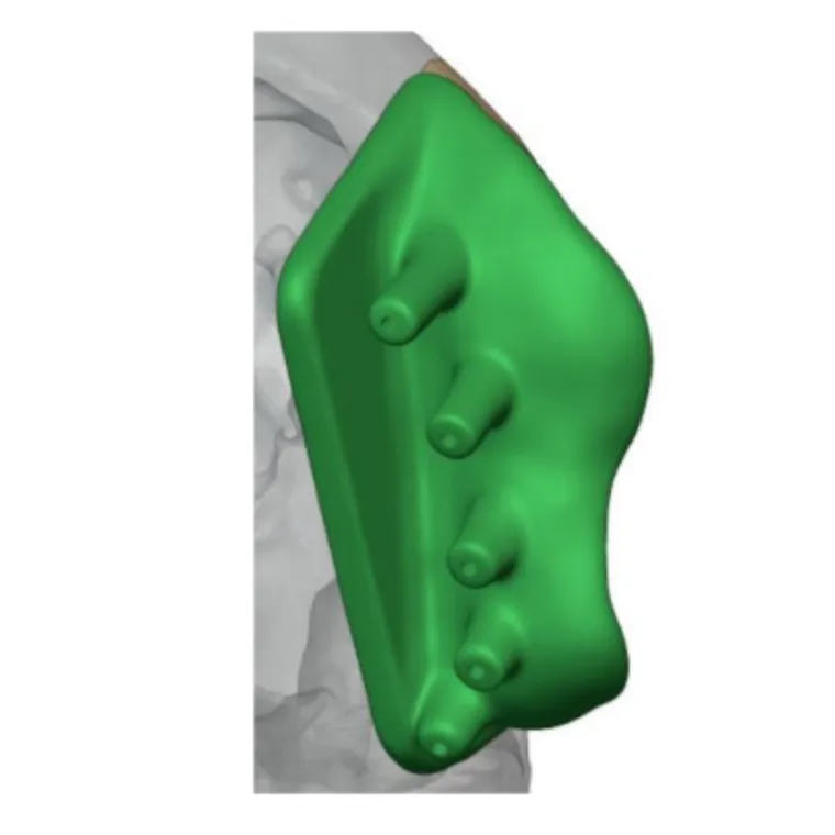
Custom implant design
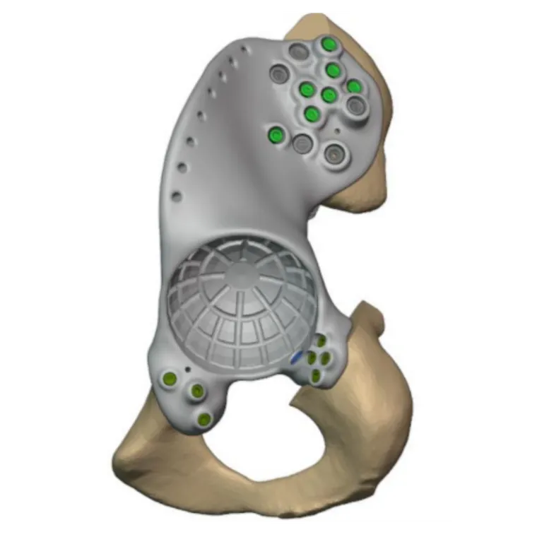
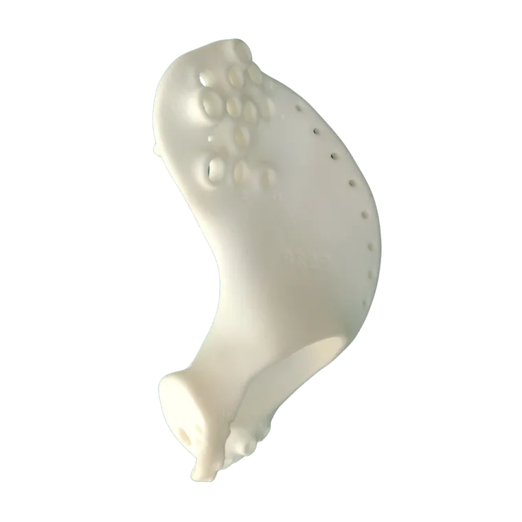
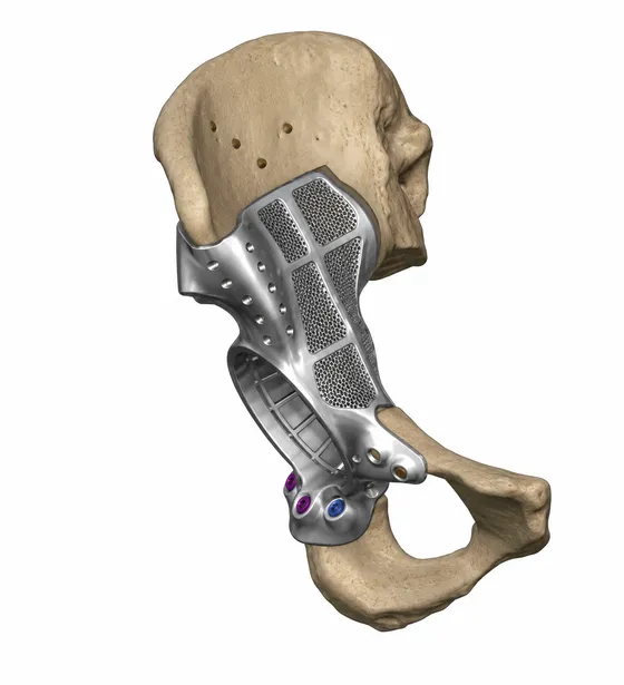
Key design features: an anatomically matched Type I + II hemipelvic implant; a porous titanium surface to promote osseointegration; a press-fit tapered iliosacral peg for primary stability; multiple locking screw trajectories into the sacrum and residual pelvis; cement-locking grooves for acetabular fixation; and compatibility with constrained total hip arthroplasty components.
Tumour resection
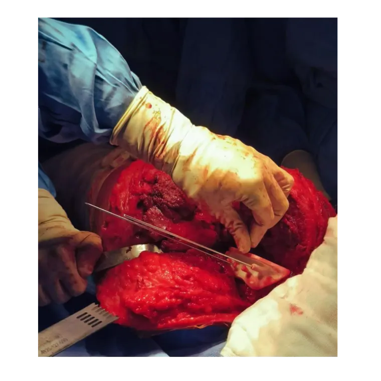
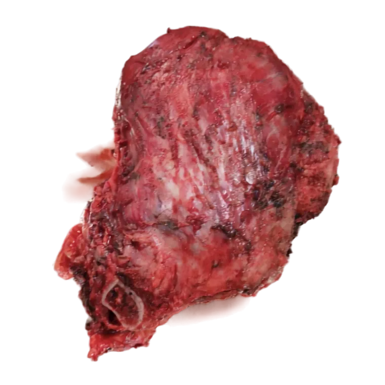
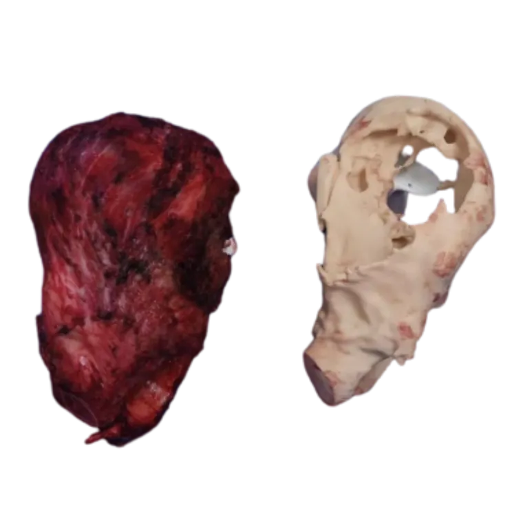
The procedure used an extended iliofemoral approach. Patient-specific cutting guides were secured with Kirschner wires, allowing precise osteotomies according to the preoperative plan. Careful dissection preserved the surrounding neurovascular structures, including the S1 nerve root. Following completion of the osteotomies, the tumour was removed en bloc.
Reconstruction
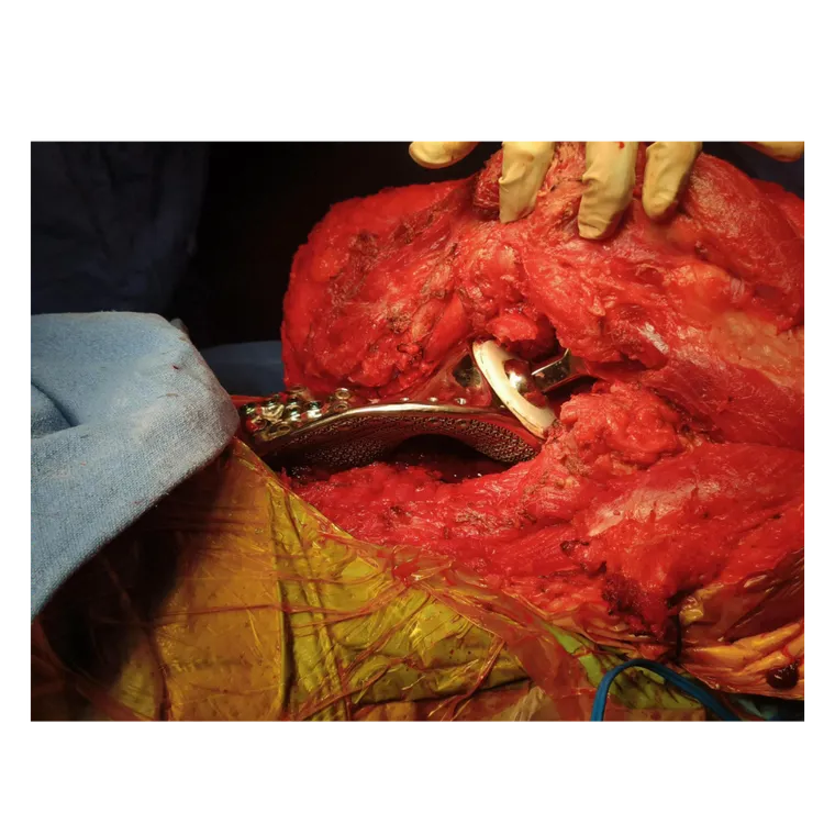
The customised implant was first trialled on the patient-specific pelvic model and native pelvis before definitive implantation. Using a patient-specific drilling guide, the iliosacral junction was prepared and a tapered porous peg inserted to achieve immediate press-fit fixation.
Final fixation was achieved with multiple locking screws into the sacrum, residual ilium, superior pubic ramus and ischium. A constrained acetabular component was cemented into the custom implant, followed by an uncemented porous-coated femoral stem and a 28 mm femoral head.
Clinical outcome
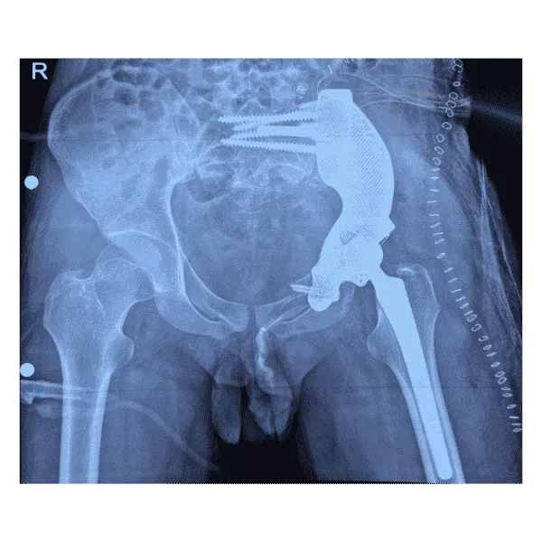
Postoperative imaging demonstrated stable implant positioning, restoration of pelvic continuity, secure iliosacral fixation, anatomical restoration of the hip centre and a stable constrained hip articulation.
Clinical significance
This case highlights the value of combining patient-specific surgical planning, customised 3D printed reconstruction and constrained total hip arthroplasty in complex pelvic tumours. The personalised approach enabled accurate tumour resection, stable reconstruction, restoration of pelvic biomechanics and rapid postoperative rehabilitation.
Related cases
 Oncology
Patient-specific FTL Cup for Tumor Resection and Acetabular Reconstruction
Pelvic tumour resection with cutting guides and a custom FTL cup
Read the case →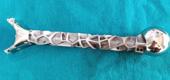 Oncology
Precision Reconstruction of Humerus with Customized Implant Solution
Custom titanium humerus prosthesis for a 10-year-old after tumour resection
Read the case →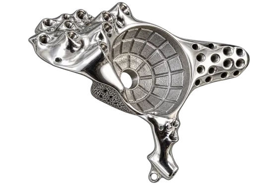 Pelvis
Pelvic Tumour Reconstruction Using Patient-Specific FTL Cup for Acetabular and Ischial Involvement
Planned resection, patient-specific jigs and custom fixation
Read the case →Have a case like this?
Send the imaging and the clinical question — first 3D visualisation back within 24 hours.