
Patient-specific FTL Cup for Tumor Resection and Acetabular Reconstruction
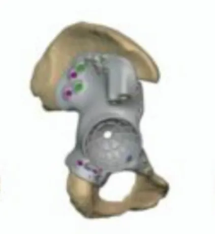
Pelvic tumour resection with cutting guides and a custom FTL cup
Patient overview
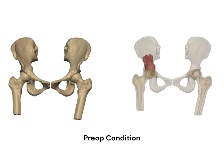
A 51-year-old female patient presented with a tumour in the right pelvis. Preoperative imaging revealed significant involvement of the acetabular region, raising concerns regarding structural integrity and postoperative joint reconstruction.
The nature and extent of the tumour were further evaluated in collaboration with the clinical engineering team to guide a precise surgical approach.
Clinical challenge
Tumour resection in the pelvic region, especially around the acetabulum, presents a significant reconstructive challenge.
Complete tumour removal required resection of a substantial portion of pelvic bone, resulting in a large acetabular defect with significant loss of the ilium. Conventional off-the-shelf implants were insufficient to restore pelvic anatomy, achieve stable fixation or support long-term joint function.
Advanced surgical planning
A patient-specific preoperative planning workflow was implemented to ensure surgical accuracy and predictability.
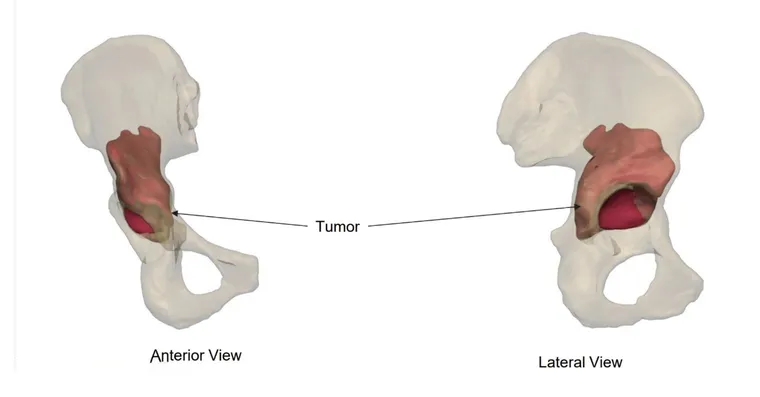
CT-based 3D segmentation enabled precise visualisation of tumour boundaries and detailed assessment of residual bone stock. Optimised resection planes were then defined to achieve complete tumour removal while preserving maximum healthy bone.
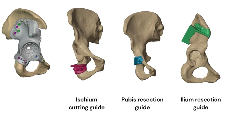
Resection with precision cutting guides
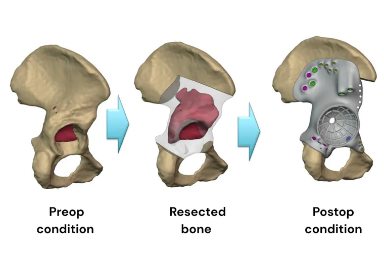
To accurately execute the surgical plan, patient-specific cutting guides (jigs) were developed. These ensure precise bone cuts along predefined planes, minimising intraoperative variability and enabling controlled tumour resection.
The jigs enhanced surgical precision, reduced intraoperative uncertainty and improved procedural efficiency.
Patient-specific FTL cup design
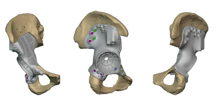
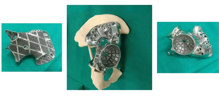
Following tumour resection, a patient-specific FTL cup was designed to reconstruct the acetabular defect and restore pelvic continuity.
The cup featured a custom-fit geometry tailored to the patient’s post-resection anatomy, ensuring accurate defect coverage and anatomical alignment.
Fixation strategy
Locking and compression screws were strategically oriented toward regions of optimal bone stock, enhancing fixation strength and long-term stability. Dedicated drilling guides enabled precise pilot hole creation, ensuring accurate screw trajectory and positioning.
The custom implant was designed to accommodate a standard cemented acetabular component, enabling reliable joint reconstruction on a patient-specific pelvic foundation. Suture holes were incorporated to facilitate soft-tissue reattachment and improve joint stability.
Postoperative outcome
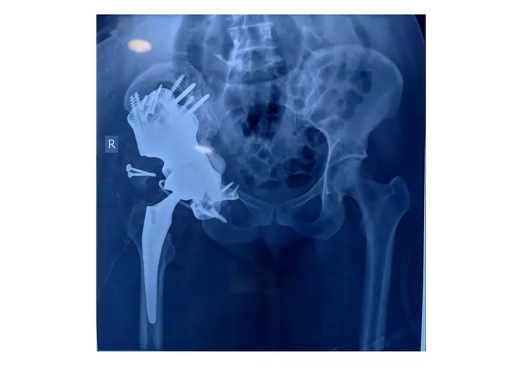
The custom pelvic implant was successfully implanted following tumour resection, achieving stable fixation and accurate reconstruction of the acetabular region.
The implant demonstrated an excellent anatomical fit, restoring pelvic structure and enabling secure placement of the cemented acetabular component. Early postoperative assessment indicated stable implant positioning and favourable conditions for functional recovery.
Clinical impact
Through advanced imaging, precise planning and customised implant design, a highly personalised solution enabled complete tumour resection, restored anatomical integrity and ensured stable reconstruction. This approach expands the scope of limb-salvage surgery in complex pelvic oncology.
Related cases
 Oncology
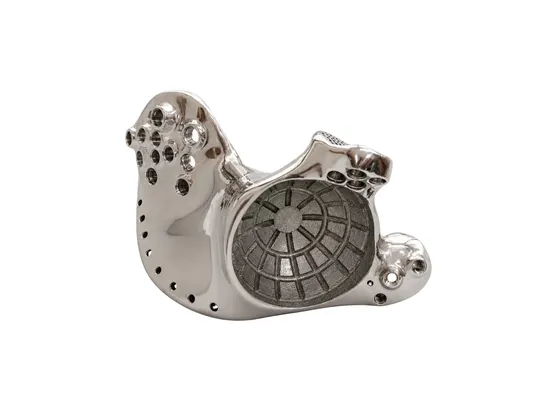
Patient-Specific 3D Printed Hemipelvic Reconstruction with Constrained THA
Type I + II internal hemipelvectomy for Ewing's sarcoma
Read the case →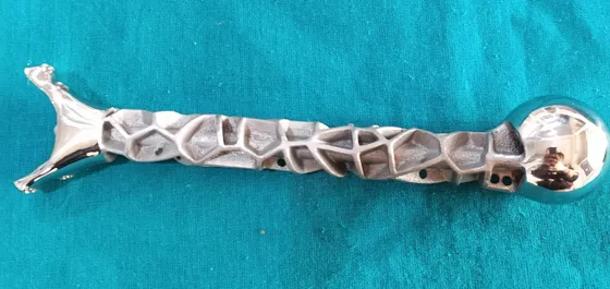 Oncology
Precision Reconstruction of Humerus with Customized Implant Solution
Custom titanium humerus prosthesis for a 10-year-old after tumour resection
Read the case →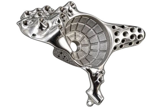 Pelvis
Pelvic Tumour Reconstruction Using Patient-Specific FTL Cup for Acetabular and Ischial Involvement
Planned resection, patient-specific jigs and custom fixation
Read the case →Have a case like this?
Send the imaging and the clinical question — first 3D visualisation back within 24 hours.