
Mandibular Reconstruction using Fibula Graft
Neo-mandible built from the patient's own fibula, planned against a mirrored healthy side
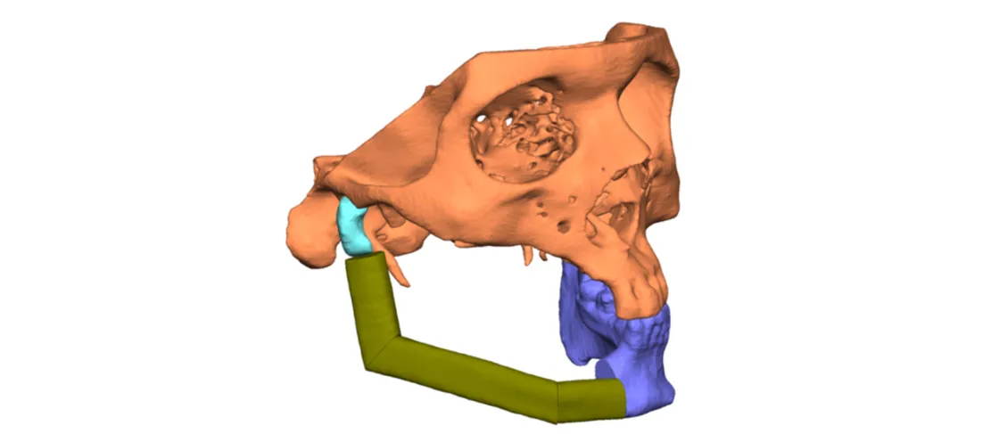
Case information
Head and neck cancer accounts for 30% of all cancers diagnosed in India. Oral cancers are the most common among males and fourth most common in females; 60-80% present at an advanced stage compared with 40% in developed countries.
There is a primary need to understand tumour location, shape and size to realise effective tumour removal followed by accurate reconstruction — either with a customised implant in bio-inert material, or using the patient’s own bone for grafting.
A male patient aged 43 was diagnosed with cancer, his primary mandible tumour removal already done. Due to the deficient right mandible he had difficulties in chewing, speech and daily activities in public because of discomforting aesthetics. Dr Raghuvir Solanki approached our clinical engineers for a robust plan and precise reconstruction of the neo-mandible using the patient’s fibula.
Visualisation
To visualise the mandibular anatomy and its neighbouring area, a high-resolution CT scan of adequate slice thickness and increment was used to 3D model the defect. The region of interest was replicated as a 1:1 digital model for understanding the defect and pathology.
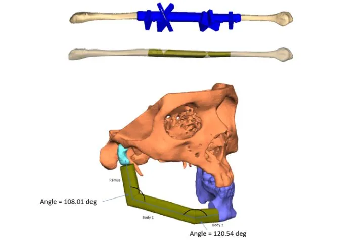
Similarly, the patient’s left fibula was modelled from the CT scan to harvest the required grafts.
Planning
Once segmentation was completed, the plan was to lay down the reconstruction of the neo-mandible using fibula bone.
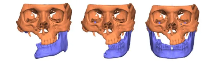
- Due to the deficient region on the right there was no contact to the right condyle, resulting in improper occlusion
- Proper occlusion was achieved by virtually simulating the correct occlusion state, taking references from the mandibular and maxillary teeth and re-positioning the healthy left condyle in the fossa
- The healthy left mandible was mirrored to achieve the best clinical reference for reconstructing the neo-mandible
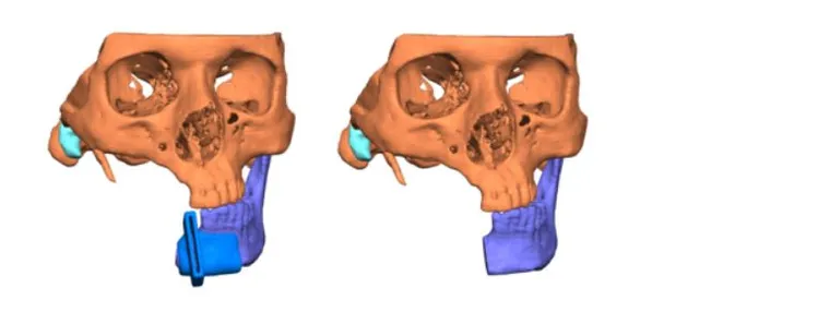
- A surgical guide was designed to resect the mandible for a perfect joint connection of the new mandible to the healthy left side
- Fibula bone reconstructed the defect following the mirrored reference, giving the right segments placed at specific angles to form the new mandible, with a surgical guide to execute the exact cuts intraoperatively
3D printing
Once the surgical plan was approved and finalised, it was translated into physical models to analyse the outcome. Pre-op, intermediate and post-op models were printed. The patient-specific guides were 3D printed in biocompatible autoclavable material for realising the planned cuts intra-operatively on the mandible and fibula bone.
Related cases
 Oncology
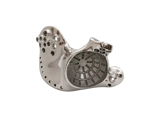
Patient-Specific 3D Printed Hemipelvic Reconstruction with Constrained THA
Type I + II internal hemipelvectomy for Ewing's sarcoma
Read the case →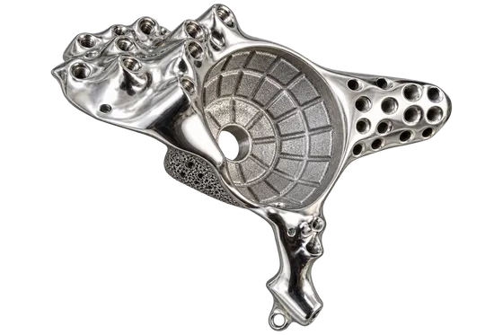 Pelvis
Pelvic Tumour Reconstruction Using Patient-Specific FTL Cup for Acetabular and Ischial Involvement
Planned resection, patient-specific jigs and custom fixation
Read the case →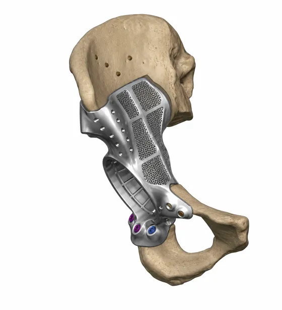 Oncology
Patient-specific FTL Cup for Tumor Resection and Acetabular Reconstruction
Pelvic tumour resection with cutting guides and a custom FTL cup
Read the case →Have a case like this?
Send the imaging and the clinical question — first 3D visualisation back within 24 hours.