
Distal Femur Replacement
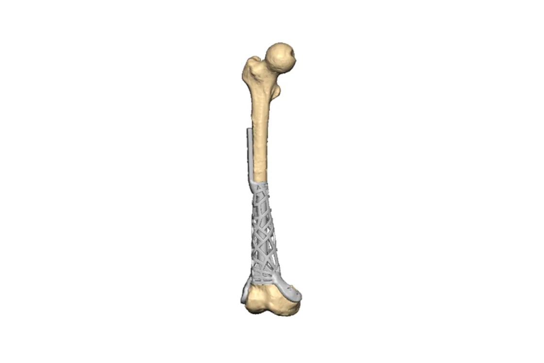
Custom intramedullary titanium implant after failed rod fixation, preserving the natural joint
Case information
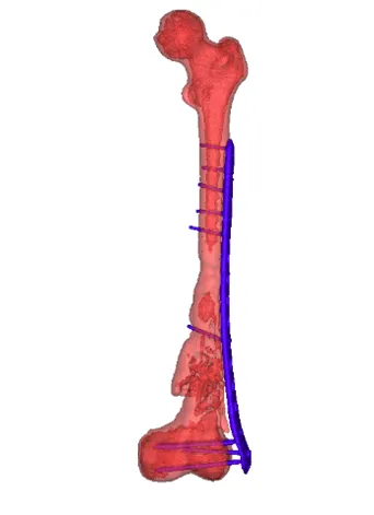
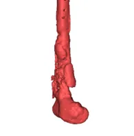
With a history of osteosarcoma 10 years ago, the patient was treated with a conventional rod implant on the femur. After a few years, the implant screws became loose and dispersed under the weight of the implant. Fragments of the distal femur were also broken, leading to severe bone damage and difficulty with mobility.
Patient-specific implants and custom guides play an immense role in the surgical approach, and a 3D model provides better case visualisation for bone reconstruction in pre-op planning.
Visualisation
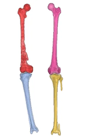
A CT scan was performed using a scan acquisition protocol. The region of interest was segmented by our imaging experts, preserving all anatomical detailing of potential clinical significance. 3D models and animations were shared with the surgeon for the initial assessment.
The initial thought was to replace the joint with an artificial implant system. However, the segmentation data showed the joint was unaffected — the distal femur and proximal tibia were intact with natural joint cartilage — so replacing the natural articulation made no sense. It was decided instead to design a customised intramedullary implant for the femur.
Planning
- Once segmentation was complete, the plan was to reconstruct the femur with a custom femur implant
- Further assessment ensured no other deformities were present
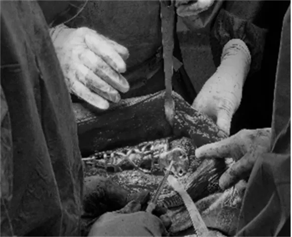
- The surgeon resected the infected femur plate using custom-designed femur cutting guides
- The healthy side was mirrored about the sagittal plane and matched to the affected limb
- Keeping a similar shape to the femur, a cage-like implant was designed with an intramedullary rod holding the femur together and flanges on the distal and proximal sides
- The resected region was reconstructed with patient-specific titanium
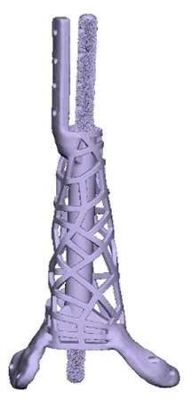
3D printing
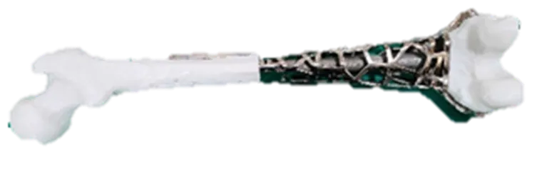
The printable plan consisted of physical 3D-printed parts — distal femur, cutting guide, drilling guide and implant prototype — which helped the surgeon evaluate the plan well before surgery.
After re-confirmation of the plan with physical models, the implants and guides were printed in biocompatible materials: titanium and medical-grade plastic respectively.
Conclusion
Pre-planning the surgical approach and using custom surgical tools and patient-specific implants all contributed to a precise reconstruction, shorter operating time and quicker patient recovery.
Related cases
 Oncology
Patient-Specific 3D Printed Hemipelvic Reconstruction with Constrained THA
Type I + II internal hemipelvectomy for Ewing's sarcoma
Read the case → Pelvis
Pelvic Tumour Reconstruction Using Patient-Specific FTL Cup for Acetabular and Ischial Involvement
Planned resection, patient-specific jigs and custom fixation
Read the case → Oncology
Patient-specific FTL Cup for Tumor Resection and Acetabular Reconstruction
Pelvic tumour resection with cutting guides and a custom FTL cup
Read the case →Have a case like this?
Send the imaging and the clinical question — first 3D visualisation back within 24 hours.