
Complex Primary Hip
Patient-specific cup positioning guide for accurate acetabular version and inclination
Introduction
Unlike other joints, the role of patient-specific instruments in THR is less explored. We took this opportunity and developed a product that could help achieve accurate acetabular cup placement. The technology can reduce error in version and inclination and give the surgeon confidence about how far they can ream. It is well documented that in cases with massive bone defects such technology is effective.
Pre-op evaluation
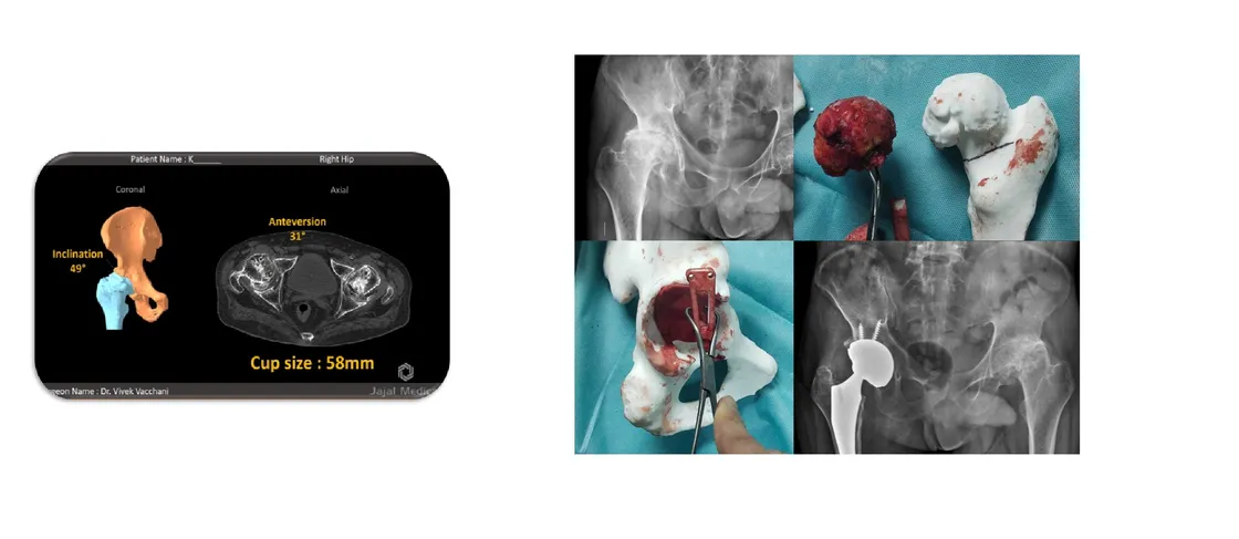
A 49-year-old male presented with pain in both hips and difficulty walking. Physical examination showed he was struggling to achieve complete range of motion, and pre-op X-rays confirmed arthritis in both hips. The surgeon reached out to Jajal Medical to explore performing this case with our cup positioning guide.
Visualise
A CT scan was performed for both hips using the scan acquisition protocol. The region of interest was segmented by our imaging experts, preserving all anatomical detailing of clinical significance. A 3D model and animation were shared with the surgeon for the initial assessment.
Plan
Dr. Vivek and our pre-op experts carried out extensive planning to ensure correct component alignment. The surgeon’s requirements were:
- A tool for referencing acetabular component alignment
- A self-explanatory pre-op model for assessment
- 3D version and inclination information
- Sizing interpretation and validation through a physical template model, including for the femur
Print and execution
The printable plan — acetabular model, component alignment guide and femur model — was dispatched in four working days, well before surgery, ensuring the surgeon had time to evaluate it. It was put into execution following a structured and cautious approach:
- Compare and evaluate the physical model against patient anatomy visually in theatre — the printed femoral head matched the actual anatomy perfectly
- After soft-tissue clean-up the cup positioning guide was placed
- Stability of the guide was confirmed before pin placement
- Feedback was taken from peer surgeons on the reference pin alignment
- Reaming was done with the reamer handle parallel to the referencing Steinmann pins
- Size prediction was evaluated by trials and confirmed against our template
- The amount of reaming required was judged against the reamed bone model
Post-op assessment
Post-op X-rays show that using this cup positioning guide, Dr. Vivek was able to decide component alignment accurately. He also shared the significance of this product for new surgeons willing to adopt a new and effective way of planning complex primary cases — it has the potential to give new surgeons confidence in the OR.
Related cases
 Hip
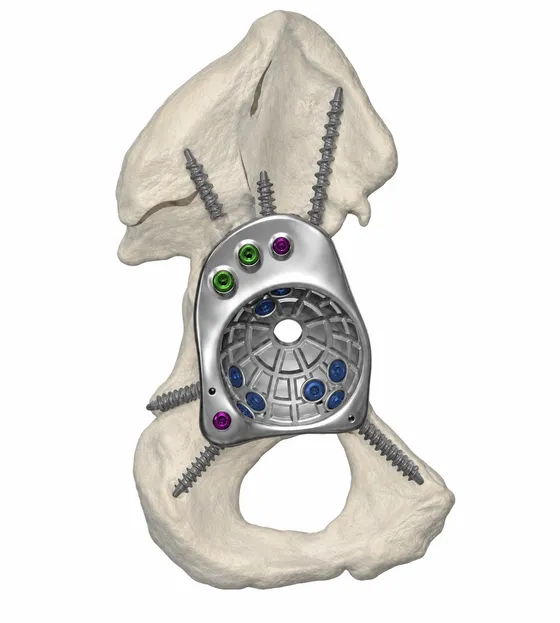
HipCustom FTL Cup for Complex Revision Hip Replacement
Massive anterior column bone loss reconstructed with a patient-specific FTL cup
Read the case →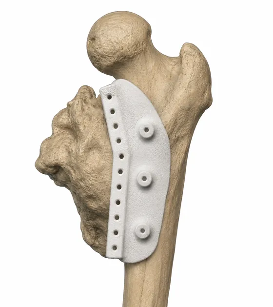 Hip
HipPrecision Resection in Heterotopic Ossification with Customized 3D Solutions
Patient-specific drilling jig planned from CT and MRI to avoid vessel and nerve injury
Read the case →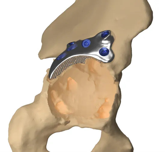 Hip
HipAdvancing Precision in Revision Hip Replacement
Custom hip columnar docking with divergent locking augment for a dislocated acetabular cup
Read the case →Have a case like this?
Send the imaging and the clinical question — first 3D visualisation back within 24 hours.