
Complex Hip Revision
Ruptured medial wall and vertical hardware shift, segmented through heavy metal artifact
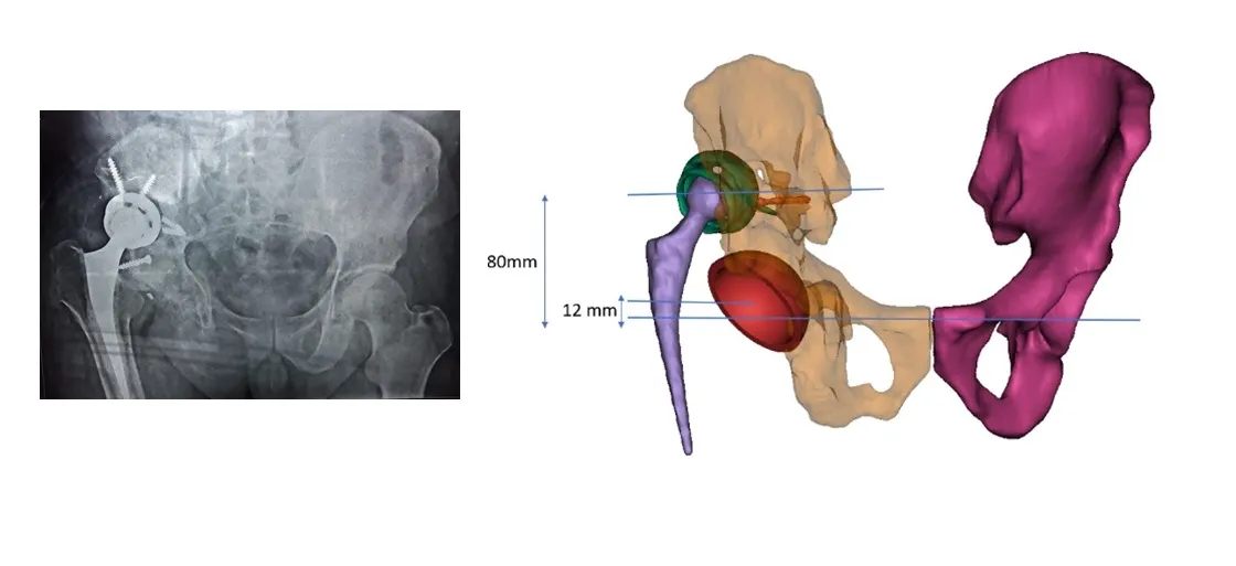
Case information
The discomfort of multiple acetabular revisions is inconceivable for the patient, and can be a nightmare for surgeons if not planned precisely. This case involved a 60-year-old male patient whose CT assessment showed a ruptured medial wall and significant vertical hardware shift.
There was a lot of artifact around the bone due to metal hardware in the CT images. Such cases need precise segmentation, avoiding artifacts and extracting the 3D model from the scan to provide the best visualisation experience.
As we move from 2D to 3D, clarity and surgeon confidence reach the next level. Our expert surgeon panel helped ensure the planning was on track, clearing all doubts around cup positioning; screw locations were re-confirmed and a robust execution plan prepared.
Visualisation
The first stepping stone to understanding the core of the problem is 3D visualisation. We used a customised segmentation process overcoming all the challenges — metal artifact, ROI identification, and preserving clinically significant regions while creating the 3D model.
Planning
The key element of the pre-op planning process is deriving clear requirements from the surgeon. Design considerations for this case:
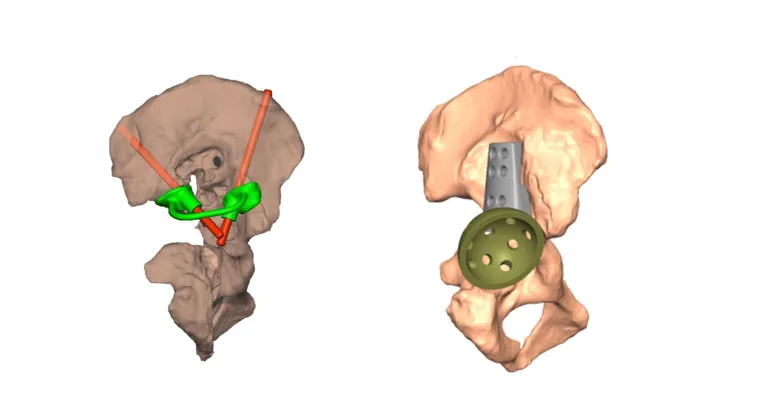
- The distorted medial wall and evident vertical displacement of hardware informed the surgeon’s decision on component placement
- Mirroring from the healthy side provided the references for cup placement
- Wall thickness analysis let the surgeon identify the denser part of the bone, helping with drilling pin location and optimising contact area
- Virtual implant positioning enabled the surgeon to decide the appropriate implant size and ensure its availability in surgery, including necessary additional augments
- Identification and creation of a patient-specific pin guide for home-run screw placement
3D printing
To raise the clinical value of the 3D-printed model we introduced the concept of Printable Planning Technology — an innovative way to engrave the physical model with all pre-op information in terms of orientation, measurements, angles and sizing, acting as a helping hand to the surgeon inside the operating theatre.
Related cases
 Hip
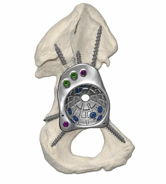
HipCustom FTL Cup for Complex Revision Hip Replacement
Massive anterior column bone loss reconstructed with a patient-specific FTL cup
Read the case →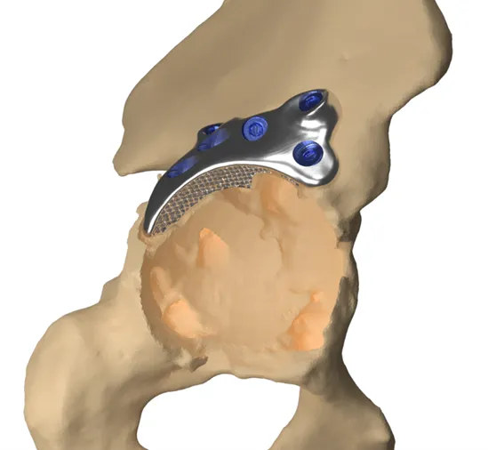 Hip
HipAdvancing Precision in Revision Hip Replacement
Custom hip columnar docking with divergent locking augment for a dislocated acetabular cup
Read the case → Revision hip
Complex Hip Revision with Severe Acetabular Bone Loss Using Patient-Specific FTL Cup
Multi-flange fixation for a deficient acetabulum
Read the case →Have a case like this?
Send the imaging and the clinical question — first 3D visualisation back within 24 hours.