

Custom Total Ankle Replacement Using a Patient-Specific 3D-Printed Implant
Patient Overview: Restoring Mobility in Advanced Ankle Arthritis
A 48-year-old female patient presented with advanced osteoarthritis of the left ankle joint, experiencing persistent pain, stiffness, and difficulty in daily movement due to severe cartilage degeneration. As the condition progressed, the ankle joint gradually lost its natural alignment and mobility, significantly affecting the patient’s quality of life.
Given the complex biomechanics of the ankle and the patient’s unique anatomy, achieving an accurate reconstruction with a conventional implant would have been challenging. To provide a more precise and long-term solution, the surgical team planned a customized total ankle replacement using a patient-specific 3D-printed implant.
Preoperative Planning: Creating a Personalized Surgical Roadmap
A successful ankle replacement depends heavily on restoring alignment, stability, and natural joint mechanics.
Working closely with Dr. Harshvardhan Hegde and his team, the Jajal Medical 3D design team carried out a detailed digital preoperative planning workflow to create a fully personalized reconstruction strategy for the patient.
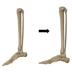
Using CT-based 3D segmentation, the engineering team developed an accurate anatomical model of the ankle joint, including the distal tibia and talus. The foot was digitally positioned in dorsiflexion along the ankle axis to recreate the patient’s normal standing alignment and restore natural biomechanics.
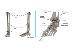
Critical anatomical landmarks and reference planes were then established to guide implant positioning and fixation planning. This collaborative planning process helped the team:
- Evaluate the deformity and joint alignment in detail
- Restore the ankle’s natural load-bearing axis
- Optimize implant positioning and fixation
- Design an implant tailored precisely to the patient’s anatomy
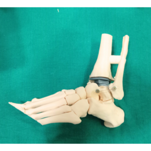
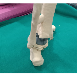
Based on this surgical plan, the Jajal Medical design and engineering team developed a fully customized total ankle implant system specifically matched the patient’s bone morphology and biomechanical requirements. First, the prototype was 3d printed and customized as per the patient’s requirements:
Patient-Specific Implant Design
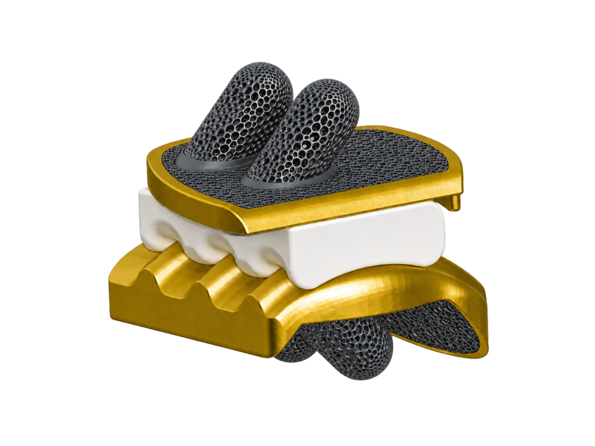
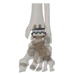
The customized ankle replacement system consisted of three components:
- Tibial Component
- Talar Component
- Polyethylene Insert (Poly)
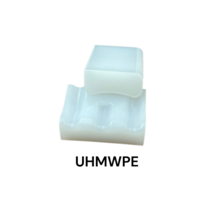
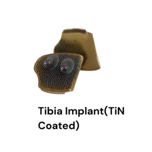
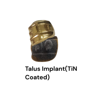
The tibial and talar components were manufactured using advanced metal 3D printing technology with medical-grade titanium alloy (Ti6Al4V), allowing highly accurate anatomical contouring along with excellent strength and biocompatibility.
The implant features patented mobile-bearing design for physiological joint mechanics, a Peg fixation interface for enhanced stability, and a Ticellium surface for ossiointegration and TiN coated articulating surface to reduce wear rate with poly. Multiple UHMWPE polyethylene insert thicknesses were available intraoperatively to facilitate patient-specific joint gap balancing and optimal implant positioning.
Surgical Execution
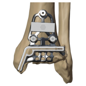
Following exposure of the ankle joint, meticulous soft tissue preparation was performed to expose the distal tibia while preserving the surrounding anatomical structures. The patient-specific distal tibial resection guide was accurately positioned on the tibia, and the planned resection level was verified using the integrated alignment wing and intraoperative C-arm imaging. Once satisfactory alignment was confirmed, the guide was secured with K-wire fixation, enabling an accurate distal tibial resection. This resection features our patented curved shape malleoli resection ensuring reduction of risk of medial malleoli fracture.
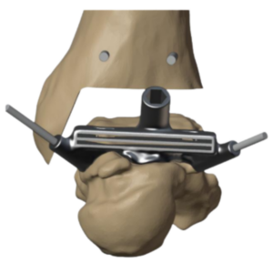
Attention was then directed to the talus, where soft tissue preparation facilitated precise placement of the patient-specific proximal talar metal resection guide. Following fluoroscopic verification of the planned resection level, the proximal talar cut was completed, after which dedicated patient-specific chamfer guides were used to perform the remaining talar resections in accordance with the preoperative surgical plan.
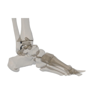
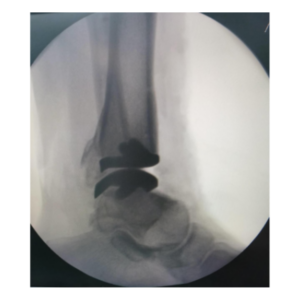
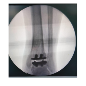
Trial components were subsequently inserted to evaluate implant fit, alignment, and joint mechanics. After confirming satisfactory positioning, peg holes were prepared in both the tibia and talus to accommodate the definitive implants. The final patient-specific tibial and talar components were implanted, with implant position verified using intraoperative C-arm imaging before insertion of the UHMWPE polyethylene insert, completing a stable, anatomically aligned ankle reconstruction.
Surgical Outcome
The customized total ankle replacement was successfully performed by Dr. Harshvardhan Hegde.
Intraoperatively, the implant demonstrated excellent anatomical conformity with the prepared tibial and talar surfaces, enabling accurate positioning and stable fixation. The patient-specific design reduced the need for excessive intraoperative adjustments and supported a smooth surgical workflow.
Postoperative assessment demonstrated:
- Accurate restoration of ankle alignment
- Stable implant fixation
- Balanced load distribution across the joint
- Improved functional recovery and mobility
Clinical Significance
This case highlights how patient-specific 3D-printed implants are reshaping complex ankle arthroplasty by enabling more personalized and anatomically accurate reconstruction strategies.
Through close collaboration between surgeon and the design team, advanced imaging, digital planning, and additive manufacturing were combined to create a highly customized implant solution tailored specifically to the patient’s anatomy.
The case demonstrates the growing value of surgeon-engineer collaboration in achieving precise reconstruction, improved biomechanical restoration, and more predictable outcomes in complex orthopedic procedures.