Club Foot Virtual Planning
Club foot is an infrequent deformity. Club foot deformity is a congenital foot condition in which the forefoot is turned inward and downward and cannot be straightened. It is also known as congenital talipes equinovarus.
A surgical approach can offer a radical form of treatment. The goal is to restore the ankle joint.
Here’s one such case of a 31yr/M patient affected with the club foot. A surgical approach can help to treat the patient. With the surgical approach better planning helps to make surgery more accessible and accurate.
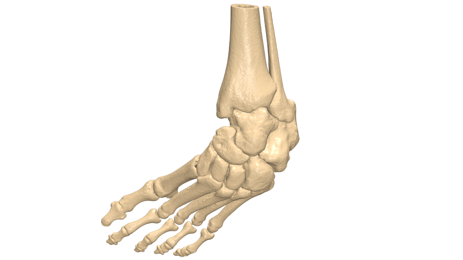
Here JMS played a vital role by virtually planning the surgery. We have mapped out the first visualization and planned the surgery. And provided a 3d pre-op model for visualization and better understanding.
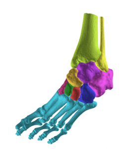
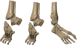
Segmentation
The region of interest was well captured by the scan and replicated in a 3d model for a better understanding and the deformity. The main goal was to provide a self-explanatory model and help the surgeon to make them visualize the information which can be missed in conventional 2D CT data.
Virtual Planning
1.The help of 3d visualization and a pre-op model provided to the surgeon that had helped the surgeon to understand and analyzed the deformity and anatomy of the club foot.
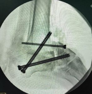
2.The Deformed Talus bone was removed. Then osteotomy was performed on the distal part of the fibula.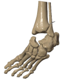
3.Osteotomy was performed on the cuboid and navicular and calcaneus bone at calcaneocuboid joint and talonavicular joint.
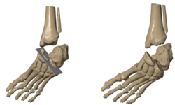
The next step was to set the position of the navicular and cuboid along with all other bones to the distal fibula at the calcaneus region.
Further, external rotation along with the medial translation and advancement was done to get the correct foot angle in line with the axis. Osteotomy was stabilized by performing the fusion using the fixation screws.
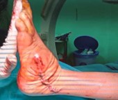
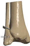
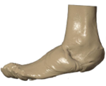
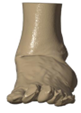
It was also required to understand the post-op soft tissue alignment and same was provided for better analysis.
Pre-op soft tissue
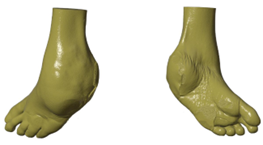
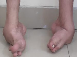
Post-op soft tissue
Conclusion
Preplanning the surgical approach and use 3d pre-op model helped in achieving accurate surgical results. And helped to achieve a straight foot angle.