3D printing for congenital heart disease (CHDs)
Ventricular septal defects
A ventricular septal defect occurs during pregnancy when the wall that grows between the two ventricles does not fully mature, producing a hole. A ventricular septal defect is one form of congenital cardiac defect. In neonates with ventricular septal defects, blood frequently flows from the left ventricle to the right ventricle and into the lungs. The excess blood pushed into the lungs strains the heart and lungs. If not corrected, this condition can eventually lead to heart failure, high blood pressure in the lungs (called pulmonary hypertension), irregular heartbeats (called arrhythmia), or stroke.
Case Information
The infant was diagnosed with VSD. During radiological analysis it was observed that the infant was suffering from the defect. The surgeon reached out to Jajal Medical for the 3d visualization before performing any surgery.
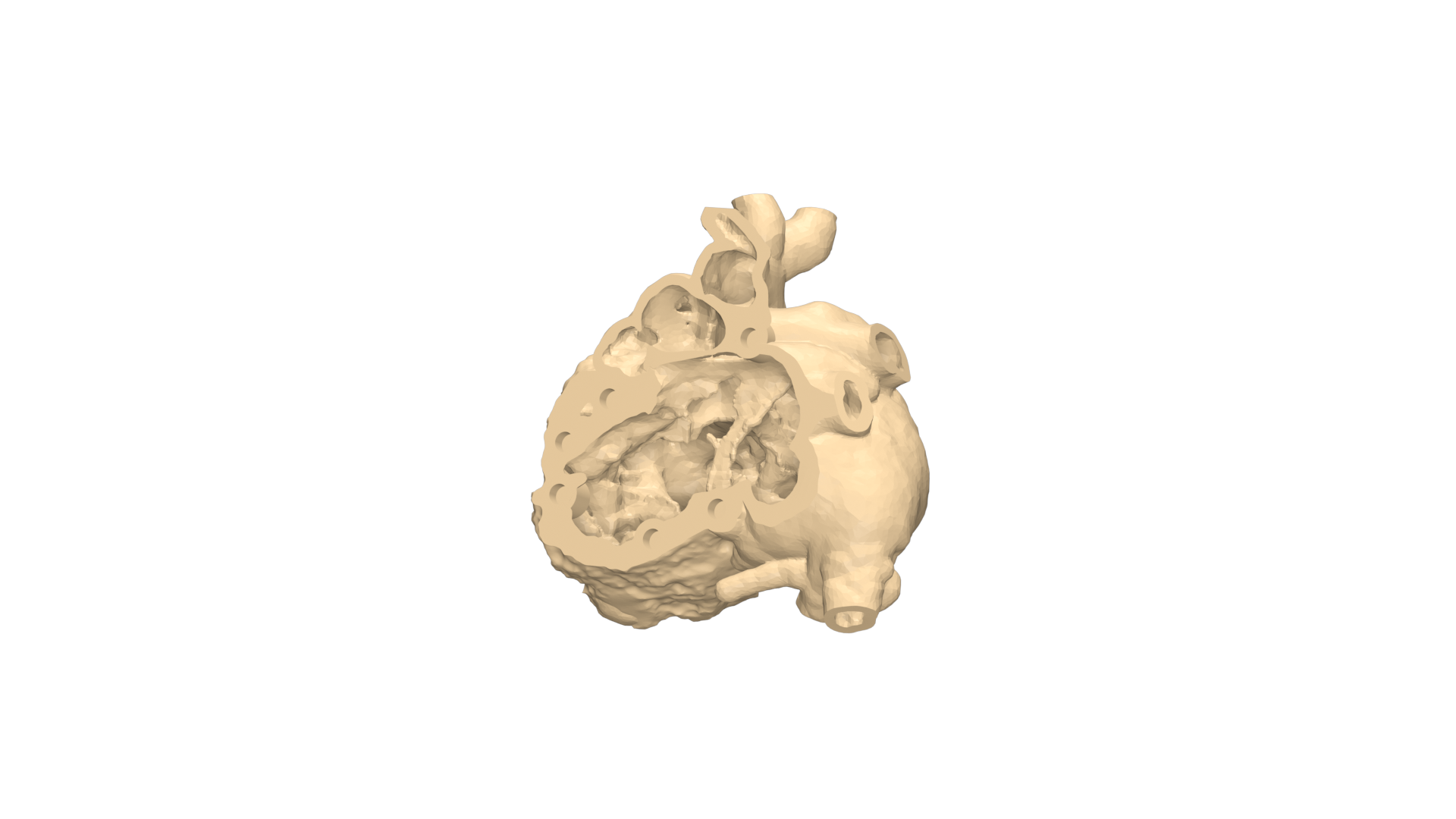
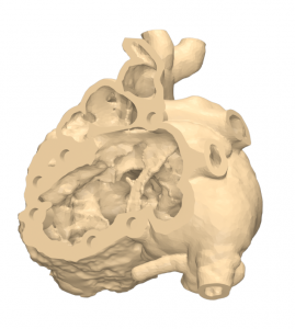
CT scan data was used with the scan acquisitions protocol. The region of interest was segmented with our imaging experts, preserving all the anatomical detailing that could have clinical significance. 3D models and animation were shared with the surgeon for the initial 3D assessment of the case.
Planning
Our clinical engineers helped the surgeon to plan as per the defect. The right patch size and the closure path were determined based on the surgical approach and the post-op results were analyzed virtually.
Printing
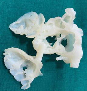
The model for plan and printed with all the accuracy and anatomical details can help the surgeon to plan the surgery and assist with a better visualization. Advanced 3D printing using photosensitive resin material was used to print the model.
Conclusion
The pre-surgical planning method, which included a custom-printed 3D model, let the surgeon visualize the procedure and make better planning decisions.